Figure 4a: Multifocal abscesses in young rat (Benny Simpson).
Case history and photos
History
Benny Simpson, a rat estimated at 8 months of age, was adopted from the humane society with 3 of his brothers. He had been at his new home for 3 weeks before presentation. Previous health history unknown.
Clinical Signs
He presented to our clinic with concerns of a fairly acutely bloated abdomen. The abdomen was distended, but not painful. A firm round mass was felt in the caudal abdomen. He was not passing a lot of stool, and the volume of urination was difficult to assess as there were frequent small amounts. Other than that, he had a 3mm raised cutaneous mass on his back. X-ray showed a granular appearance of the abdominal contents, with very poor detail to the abdomen. The chest cavity was normal on X-ray. Ultrasound showed a 3.8 cm circular mass. It was very homogeneous and isoechoic to the liver parenchyma. This was not consistent with a fluid filled cavity (i.e., blocked bladder). The attempt at ultrasound examination was extremely limited as Benny was not sedated, and seemed uncomfortable for positioning, thus only a few seconds of scanning was done. As the mass was interfering with his ability to defecate normally, and seemed to be compressing his abdominal organs somewhat, the decision was made to do an exploratory surgery the next day.
The following day Benny was still bright, active, eating, but seemed somewhat more distended. He was anesthetized. While under anesthetic another brief ultrasound check was done (we didn’t want him under anesthetic too long as the mass would potentially compress blood return). At that point we saw the same mass, now slightly larger, but 2 other round masses were also seen near it. A ventral midline incision was made. There were very small encapsulated growths incorporated into the linea alba, and there were adhesions of the omentum to the body wall (non-medically-there were very small abscesses in the central ligament that is opened in the abdomen, and the coating structures of the abdomen were stuck to the body wall). There were a chain of spherical masses from the midline body wall, 1.0 – 1.5 cm encapsulated masses, also dozens of 3-4 mm inclusions in the omentum, and around the bowel. In the mid-caudal abdomen, the 4.5 cm discrete mass was present but it was adhesed to the body wall, and had bowel adhesed to it (see photos below). The liver had 4 masses presented incorporated into the parenchyma involving >50% of the organ. The spleen had 2 growths, and each kidney was >70% of the parenchyma involved with the masses. Based on the extensiveness of the lesions (50-60 discrete growths), the progression of the condition even over 24 hours, and the fact that every major abdominal organ was involved, the condition was deemed inoperable.
Diagnosis
The working diagnosis was a previous septicemia (bacteria in the blood stream) seeding the abdominal cavity, vs a very unusual cancer spread.
Treatment
N/A
Outcome
Benny was euthanized while under anesthetic, and samples were taken for pathology and culture & sensitivity due to multiple siblings present.
Follow-up
Histopathology demonstrated that the masses were indeed encapsulated abscesses with degenerated neutrophils (white blood cells), debris and large bacteria colonies, surrounded by a thick fibrous tissue capsule. The bacteria colonies were filamentous, gram positive rods that with further testing did stain with acid fast stain. Microbiology grew a large population of beta-hemolytic streptococcus lancefield group G. The filamentous Gram-positive rods were observed as well, but did not grow in culture, even with extended incubation. The differentials for this bacteria were nocardia species (pathology was leaning towards this), or actinomyces. Based on the lack of soil exposure, and the inability of this bacteria to grow in the lab environment, the likely diagnosis of actinomyces was made (actinomyces is generally very difficult to isolate). This organism is normally present in the cheek of rats, and definitely could have caused a generalized septicemia.
Discussion There still remains unanswered questions in this case. The main one for me was how was Benny so bright and active, when about 30% of his body weight was abscess and infected tissue? Also, how was he physically able to grow these abscesses so rapidly? The abdominal swelling was noticed over a period of a couple of days at home, and we saw a noticeable change in 24 hours at our clinic. We will also not know when the initial septicemic episode occurred.
Photos
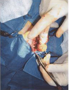 Photo 1: Taken during surgery. |
 Photo 2: Taken during surgery showing grape-sized abscess. |
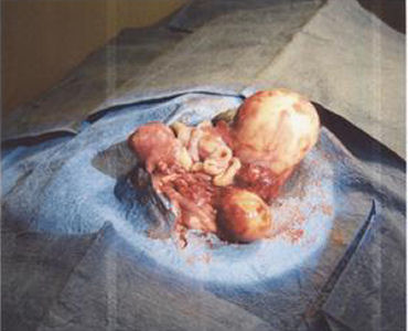 Photo 3: Shows tangerine-sized abscess taken post-mortum. |
Case history and photos courtesy of:
Dr. Mimi Ehrlich
Ajax Animal Hospital
Ajax, Ontario Canada
http://www.ajaxanimalhospital.com/

