Figure 5b: Scrotal abscess in 9-month-old male rat
Case history and photos
History
Osmolarity, 9 months old, ~500 grams, male, in a 4′ X 6′ Midwest Ferret Cage with 7 other furry gentlemen ranging in age between 6 months and 1.2 years of age.
Clinical signs
Extreme scrotal swelling and bruising. Rectal bleeding. Bowel obstruction. Discomfort.
Diagnosis
Possible Granuloma.
Possible abscess.
Treatment
2 injections of BiPen (Benzathine Pen/Procaine Pen G.) 30,000 IU/kg bwt. administered every 6 days.
Follow up
Right before Christmas swelling became more noticeable in Osmo’s testicles occurring overnight. I never noticed the bite wound that started it all. My vet didn’t see it either.
After the trip to the vet’s, I gave Osmo another salt water enema to help clean him out (apparently it was too painful to actually pass stool on his own) and peered in with an otoscope. To the right I saw the infected mass which looked to be about 1.5 cm in diameter. Surrounding it was a lot of raw looking tissue, some of which was
lined with a less granular, greenish color infection. Looking again outside, I was able to see a scabbed-over hole where the wound originated.
Within 24 hours following the administration of the BiPen injection, the swelling was greatly reduced to the point where Osmo no longer required assistance nor tolerated interference in helping to evacuate the bowel of stool. After 5 days, very little was left of the infection itself, the swelling was completely gone, and the bite wound that caused the severe abscess was now seeping and scabbing over.
Within 24 hours of the administration of the second BiPen injection on the 6th day, the infection was no longer visible at all.
Outcome
Healed.
Photos
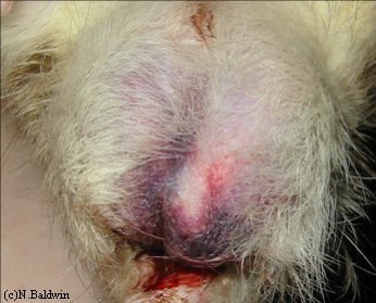 Photo 1: Significant discoloration/bruising of the testicles and a small amount of rectal bleed. Photo taken before treatment. |
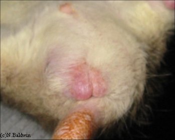 Photo 2: For comparison, normal view (note: taken in cool weather) from below. Photo taken approx. 2 weeks after initial treatment. |
 Photo 3: Front view of bruising and bleeding. Photo taken before treatment. |
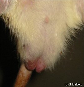 Photo 4: For comparison, normal frontal view. Photo taken approx. 2 weeks after initial treatment. |
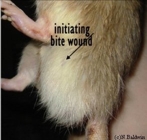 Photo 5: Showing presence of bite wound. Photo taken 6 hours after first BiPen injection. |
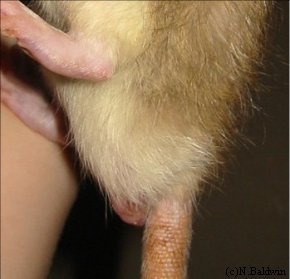 Photo 6: For comparison, normal side view (note: taken in cool weather). Photo taken approx. 2 weeks after initial treatment. |
Case history and photos courtesy of Nathalie Baldwin at RatRaisins,Inc

