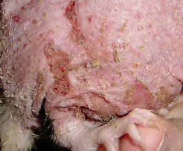
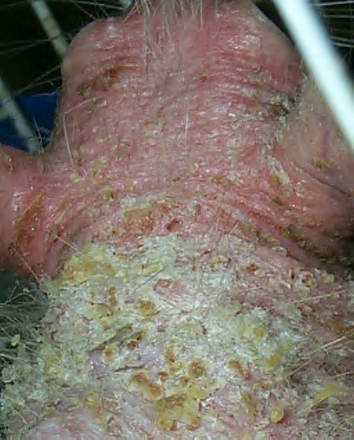
Figure 1: Pyoderma in female rat (Ruby).
Case history and photos
History
Ruby is an adult female rat.
Clinical Signs
Presented with crusted lesions and inflammation of the epidermis and hair loss.
When the usual treatments for dermatitis didn’t work, Ruby was started on 3 weeks of Ivermectin to treat a presumed parasitic infection. When this too failed, she was placed on azithromycin for a week in combination with prednisone (anti-inflammatory). It helped but didn’t resolve the problem. After getting worse (skin was dry and swollen) we returned to the prednisone and targeted a more topical approach covering a variety of antibacterial salves. Having no luck with that, we took the advice of Dr. Morrisey at AMC who recommended getting a biopsy on one of her lesions:
“Submitted is a skin biopsy, and examined are multiple sections. In this skin biopsy, there are multifocal
ulcerations and erosions of the overlying epithelium. These erosions and ulcerations are covered with a closely
adherent serocellular inflammatory crust. Large numbers of neutrophils are infiltrating across areas with intact
epithelium. The neutrophils are also present in the superficial dermis. In these sections, there is reduction
in size of the follicles as well as numbers. In areas, they are completely effaced by the inflammation. Small
clusters of bacteria are identified within the crust. These bacteria are coccoid shaped organisms.”
Diagnosis
Results from the biopsy:
“Severe diffuse erosive and ulcerative pleocellular dermatitis with crust formation and intralesional bacteria.”
“The skin lesions are primarily of an ulcerative inflammatory dermatitis. The bacterial organisms identified within the crusts are suggestive of possible staphylococcal organisms. They are less likely streptococcus.
The lesions are usually concentrated in the interscapular region and dorsal thoracic areas in rats. Staphylococcus aureus is the most common isolate. As self-induced trauma plays a role in the disease development, toenail clipping of the hind feet of affected rats can greatly reduced the severity of lesions.
The prominent lymphocytic as well as plasmacytic component also suggests a hypersensitivity reaction. This may be in response to the bacteria or environmental antigens.”
Treatment
Suggested treatment for the staph infection:
Three weeks of antibiotics using a cephalosporin, twice a week bathing with oxydex shampoo.
Prognosis
According to Dr. Morrisey, with treatment the lesions would cease to spread, begin to heal, and the fur to grow back.
Outcome
Unfortunately Ruby was already very ill, and succumbed soon after the final treatment began.
Photos
|
 |
|
|
Photos 1 & 2: Serous crusted, erosive and ulcerated lesions with hair loss can be seen in both photos. |
||
Case history and photos courtesy of Nathalie Baldwin at RatRaisins,Inc

