Figure 5g: Ovarian abscess in an intact female doe. (unnamed)
Case history and photos
History
Intact Doe, of unknown age (found as a stray), BIOP 7m. Owner: N. Woolmer RVN. Owner palpated an abdominal mass. An ultrasound was scheduled for the following day.
Clinical Signs
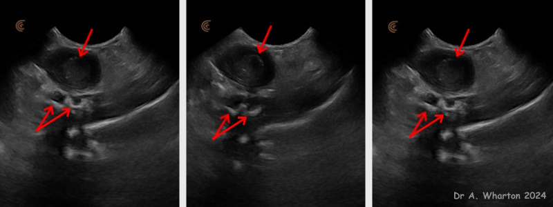
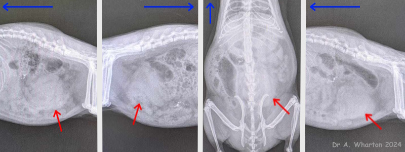
Ultrasound showed what appeared to be a soft tissue mass within the bladder. However, it didn’t look typical for a transitional cell carcinoma – it was very round, smooth and homogenous in appearance. Fluid could be seen in two uterus horns, which did not make sense unless there were 2 separate issues. The mass didn’t look dense enough to be a urolith, and there was no acoustic shadowing, but to be sure xray was performed on the following day under iso/O2. Xray suggested it might not be a bladder mass as bladder is normal size and radiodensity, and the mass can be seen in ventral abdomen.
Diagnosis
Abdominal mass. Discussed with the owner and decision was made to do exploratory laparotomy.
Treatment
Ex lap schedules. The doe was well in herself, so surgery was performed 5 days after the discovery of the mass. The doe was given methadone/ketamine premed, followed by induction and maintenance with iso/O2.
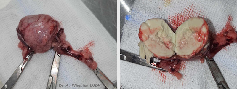
She had oral meloxicam that morning, and maropitant was given subcutaneously. Fluids were administered throughout. A single midline laparotomy incision was made, and the mass located. On exteriorising the mass, it appeared to be a neoplasm of the left ovary, wrapped in omentum. The uterine body contained a little fluid. The cervix and the right ovary appeared normal. Ovariohysterectomy was performed, with autoconstricting pga ligatures on the pedicles and cervix. A Standard midline closure was performed, with a lidocaine block to the incision, and intradermal sutures in the skin. Recovery was uneventful.
Outcome
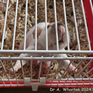
The doe felt so well and was So Very active she returned with a hernia from having popped a muscle suture. This was treated and she continues to do well.
Photos

The first photo shows the single red arrows indicate what was thought to be the soft tissue mass within the bladder. Fluid can also be seen in two uterus horns (indicated by double red arrows), which didn’t make sense unless there were 2 separate issues. The mass didn’t look dense enough to be a urolith, and there was no acoustic shadowing, but just to be sure she was x-rayed the following day under iso/O2. |

Photo shows that x-ray images suggested it might not be a bladder mass – the bladder is normal size and radiodensity, and the mass (indicated by red arrows) can be seen in the ventral abdomen. The blue arrows indicate the cranial direction. |

Photo post surgery: the ovarian mass was incised, and found to be an abscess containing thick, odorless pus, explaining the homogeneous appearance on ultrasound. |

Photo shows female rat recovered from post op. |
Owner: N. Woolmer RVN
Case history and photos courtesy of Adele Wharton, BVSc, MRCVS, CertGP
Case and photo compilation by Cyzahhe
Case editing courtesy of Karen Grant RN and Cyzahhe


