Figure 11: Postop splenectomy of grossly enlarged spleen showing abnormal structure in male rat (Obi Foster)
Case history and photos
History
Obi Foster, 7-month-old intact buck. From NFRS registered breeder and been with owner from 8 weeks old, no previous health concerns.
Clinical Signs
Presented for a routine health check, and on abdominal palpation his spleen was found to be grossly enlarged. He was reported to be showing no clinical signs at the time.
Diagnosis
Ultrasound confirmed abnormal internal structure of the spleen, particularly at the head, which were suspected to be neoplastic changes.
Plan of action
After discussing the findings with his owner, and that no obvious signs of metastatic disease were seen on ultrasound (though advising that this did not exclude microscopic metastasis) he was scheduled for splenectomy.
Treatment
On the day of surgery, he remained clinically well, although possibly a little uncomfortable.
He was given a methadone/ketamine premed and induced and maintained with sevoflurane in O2. He was given meloxicam, maropitant to reduce postop nausea and inappetence in the rat, as well as TXA (tranexamic acid to help with any clotting issues that might arise) before surgery began.
Splenectomy was performed via a ventral midline incision, and constrictor ligatures placed on the splenic vessels. The spleen itself was grossly normal in external appearance, with only 2 small nodules present, but 3 – 4 times the normal size. There was no visible sign of metastasis.
Standard midline closure with a lidocaine block, and intradermal sutures in the skin.
Recovery was uneventful, although he did chew a suture in the immediate postoperative period which required repair with tissue glue.
Follow-up
Histopathology was not performed on the spleen, but the organ has been preserved in formalin for later submission if Obi develops any potentially related problems.
Outcome
Obi Foster is continuing to do well to date.
Photos
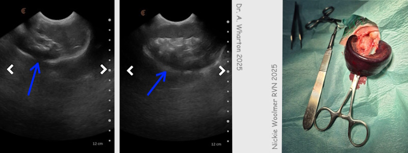
The photo on the left and center with blue arrows shows spleen. The photo on the right shows removed, enlarged, spleen with scalpel next to it for scale (size comparison). |
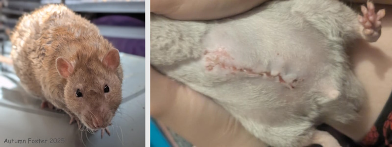
The photo on the left shows Obi prior to surgery. The photo on the right shows incisional healing. |
Case history and ultrasound images courtesy of Adele Wharton, BVSc, MRCVS, CertGP
Photo of spleen with scalpel for scale courtesy of Nickie Woolmer, RVN
Photos of Obi prior to surgery and healing courtesy of Autumn Foster, owner
Photo compilation by Cyzahhe
Case editing courtesy of Karen Grant RN

