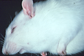
Figure 2: Coronaviruses (RCV and SDAV)
photos courtesy of IDEXX RADIL
A. Etiology: Coronavirus is an RNA virus with 2 strains identified to cause disease in rats. Rat coronavirus (RCV) causes respiratory infection while sialodacryoadenitis virus (SDAV) infects the upper respiratory tract, Hardarian and exorbital lacrimal glands, and the submandibular and parotid salivary glands. The SDAV strain causes clinical disease.
B. Transmission: RCV/SDAV is highly contagious and is spread by aerosol, direct contact, and fomites. No latent infection or carrier state occurs. The incidence of infection is high.
|
C. Clinical Signs: Viral infection is not fatal, and is generally subclinical. Rats infected with SDAV may exhibit a porphyrin oculonasal discharge. The submandibular salivary gland may be palpably enlarged due to sialoadenitis (see photo). Dacryoadenitis may cause exophthalmos, which can lead to keratitis and corneal ulcers. Symptomatic rats are at a greater risk for inhalation anesthesia. Chronic exophthalmos can result in exposure keratitis.
|
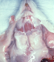
D. Pathology: Lesions include enlarged submandibular and parotid salivary glands, edematous cervical lymph nodes, swollen lacrimal glands, and yellow-grey foci in Harderian glands (brown-red mottling of the Harderian gland is normal due to porphyrin production).
Histopathological examination of submandibular salivary glands reveals degenerative changes including acinar and ductal epithelial necrosis, interstitial edema, squamous metaplasia of ductular epithelium followed by proliferation of hyperchromatic acinar cells. Chronic lesions include replacement of acini with fibrous connective tissue and an infiltration of lymphocytes. Similar lesions are present in the lacrimal and Harderian glands. The sublingual salivary gland (mucous gland) is usually not affected.
E. Diagnosis: The most common diagnostic test is serologic screen for antibody and include ELISA and immunofluorescent antibody assays. Coronavirus PCR assays will help detect RCV or SDAV in acute infections. SDAV PCR samples include Hardarian gland or submandibular salivary gland, and while lung is the optimal RCV sample.
F. Control: No carrier state exists, and recovered rats are free of virus and are immune. Cessation of all breeding activity in a production colony for 60 days and concomitant removal of all suckling and weanling animals or intentional exposure of these young rats by frequent mixing will eliminate the infection in the colony. The immune status of the colony and of any new rats to be introduced should be monitored serologically.
Special thanks to the University of Missouri – Comparative Medicine Program and IDEXX-BioAnalytics, and to Craig Franklin, DVM, PhD Associate Professor, for allowing us to use this information on the Rat Guide.

