Figure 4: Squamous carcinoma of the inner ear (with ulceration, abscess, and resulting inner ear infection) in female rat (Jet).
Case history and photos
December 21, 2002:
Celeste (“Jet”), spayed female black Berkshire age 26 months (347g), presents with reddish-brown waxy discharge in her left ear. She cleans it herself. No other symptoms.
December 22, 2002 – January 13, 2003
During this time, Jet presents twice more with the same discharge noted above. Again, she cleans it herself. No other symptoms.
January 14, 2003
Jet presents with moderate swelling just under her left ear and mild head-shaking. Reddish-brown discharge visible inside the left ear as noted above. No other symptoms. Made a next-day appointment with the general care vet.
January 15, 2003
Reddish-brown discharge and head-shaking the same, swelling unchanged.
New symptom: Bruxing now seems to bother her. General vet swabbed ear, checked for mites (note: rats are not susceptible to mites in the ear canal) and palpated the swelling.
Dx: Swelling of undetermined nature.
Rx: Amoxil .05mL SID, Otomax ointment BID, swab ear BID, Baytril 5mg SID. (Note: Fine needle aspiration of the swelling would have been in order.)
January 16, 2003
New discharges: White liquid now visible in ear canal, porphyrin staining in left nostril. Right eye appears to protrude (note: This symptom could be confused with a sunken left eye, common to many ear infections). Took Jet to the general care vet who swabbed the area believing the white substance to be “ear wax” (more probable is a bacterial infection).
Rx: Continue treatment as above and recheck in 7 days.
Later that evening husband says he’s been smelling a mild foul odor from her ear for several days (I have no sense of smell) which would indicate a bacterial infection.
January 17, 2003
After two days of treatment, no change in swelling. Ear presents with whitish discharge with what appears to be white beads suspended in it. Head-shaking and interrupted bruxing persist; no other symptoms observed. Called for appointment with an exotic specialist in our area, only available time slot was for Tuesday, January 21st, told to call again the following day. Later in the evening, the discharge takes on a greenish cast.
 Photo 1: 1/17/03 Ear discharge can be seen. |
January 18, 2003
Appointment with exotic specialist Michael Doolen, DVM, of Oahkhurst Animal Hospital. Described Jet’s history, the current meds and treatment, and the current symptoms. By this time, all discharge in the ear canal is whitish-green, no reddish-brown discharge at all. Mild head-shaking persists; swelling is somewhat worsened.
The Vet anesthetized Jet with ISO, obtained a swab of the discharge, and performed an otoscopic evaluation along with a fine needle aspiration of the swelling. Swab revealed exudate consistent with a 2nd degree bacterial infection. Visual exam revealed a red mass in the ear canal, possibly blocking the lower ear canal completely. Fine needle aspiration revealed a few RBC’s and some rare large round cells in the lower ear canal consistent with a cancerous mass.
Dx: Ulcerated neoplastic mass in ear canal with second degree bacterial infection.
Rx: Continue Otomax daily for 7 days, flush ear daily with Acetic Acid rinse, swab excess matter from the ear canal. Discontinue Amoxil but continue Baytril at half the dosage (2.5mg SID). Depomedrol .04mL injection performed on-site, additional injections will be needed every 10-14 days.
Because the mass and the area in which it was located were both highly vascular – and also due to Jet’s age – reducing the size of the tumor surgically was ruled out for the time being.
January 22, 2003
Head-shaking persists, discharge has lessened. Swelling has increased and taken on a purplish color below the ear, on the cheek area. Skin in that area is shiny and stretched. Jet is still very active and otherwise fairly asymptomatic, appetite fairly good but Jet has always had a lean physique and been a picky eater. Weight: approx 336g (converted from home portion scale, accurate in oz to one decimal place).
January 23, 2003
Swelling has increased approximately 60% since yesterday, consistent with large abscess. Discharge continues to lessen. Vet will do exploratory examination on 1/25. Possibility of surgery discussed, also cauterization if possible and/or necessary.
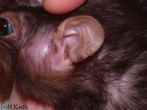 Photo 2: Abscessed mass and swelling, 1/23/03 |
 Photo 3: Abscess mass with scab (pre-surgery), 1/24/03 |
January 25, 2003
Vet administers ISO and opens the skin over the swelling. Large amount of pus removed.
Dx: Lavaged abscess. Opening the abscess reveals that the size of the neoplastic mass is unchanged. Vet cauterized the mass with radiosurgery and installed a drain to keep the wound open and flushable.
Rx: Penicillin .05ml (injectible) SID and Chlorhexidine to flush twice a day. Continue with Baytril.
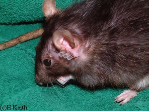 Photo 4: Post-op drains for abscessed mass, 1/25/03 |
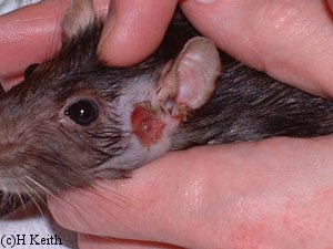 Photo 5: Opened abscess (freshly flushed), 1/27/03 |
Late January – February
Though the drain was sutured into Jet’s skin, she managed to shed it within the next 24 hours. The skin around the wound simply failed and sloughed off completely, leaving the wound completely open and crater-like. We swabbed the area very gently until the skin had healed from the radiosurgery.
After the initial healing period, we began to flush aggressively with Chlorhexidine through a needle-less syringe in order to keep the wound open. The abscess wound was complicated by white blood cell exudate, which presents as a firm white wax, coming from a very small opening that led from the wound directly into her ear canal (see photo). While the outer infection would resolve in time, the inner irritation and resulting infection never did.
Once the wound closed, the infection in her lower ear canal abscessed again.
 Photo 6: Shows drain sutured to skin. |
 Photo 7: Following drain removal |
 Photo 8: Improvement noted with flushing of wound. |
 Photo 9: Site of abscess appeared to be somewhat improved, however swelling persists. |
March 22, 2003
With the side of her face swollen up, we took Jet to the vet. He injected the swelling with lidocaine and lanced the abscess. There was a lot of caseous debris (milky green fluid and hard white waxy substance) which he flushed out with a needle-less syringe. We decided there was no choice; we scheduled a lateral ear canal resection for the following week to permanently open Jet’s ear canal and prevent further complications.
March 24, 2003
Lateral ear canal resection performed under ISO anesthesia. Opening the area revealed that the tumor had grown very large and had completely filled the middle ear. Vet removed about three cubic mm of the tumor, but the tumor had invaded very deep and complete removal was not possible without invading too close to the skull and TMJ (temporal mandibular joint, ie., jaw). Vet cauterized remaining tissue.
Dx: Vet’s notes indicates the tumor has “the appearance of SCC” (squamous cell carcinoma), which we had previously discussed as a likelihood.
Rx: Leave the surgery site completely alone for two days. Interferon injection (.02ml) given on-site with supply to take home and administer once every three days.
March 25 – 30, 2003
Jet tolerated the major surgery fairly well. There was some bleeding and the infection was still in the ear canal, but after a couple days all symptoms began to resolve. During playtime later in the week, Jet took a few missteps. (We would later learn this was due to the tumor invading her brainstem.) The next evening, I noticed Jet was having some respiratory distress. Fearing a myco flareup, we steamed her in the bathroom, set a humidifier to steam into her cage overnight, and planned to made an appointment to see the vet the next day.
March 31, 2003
I called from work to make an appointment. Based on my description of her symptoms, the vet requested to see her right away. When I arrived home later in the morning to pick her up, Jet was awake (unusual for her in the daytime) and apparently in distress. Her eyes were very wide, she was rolling severely, and she could not keep her balance. I placed her into a carrier and rushed her to the vet.
Upon seeing her symptoms, the vet informed me there were no further treatment options available. Either the infection had spread through to the other ear, or the tumor had invaded her brain. Having no option left, we chose to have Jet euthanized.
April 1, 2003
Necropsy revealed that the mass was indeed squamous cell carcinoma that had invaded her brainstem medially. No other abnormalities were noted aside from the remaining infection in her ear canal, which was on its way to resolving.
Photos showing Jet’s supplies for flushing of wound can be accessed here: Fig. 4b Jet’s Supplies.
Case history and photos courtesy of H. Keith at: http://www.propellertail.com/scc1.htm

