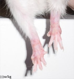
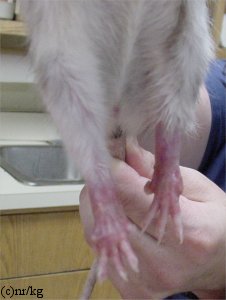
Figure 2: Pyoderma of the hind limbs
Case history and photos
History
Adult female rat brought with cage mate to Alamo Veterinary Clinic when owners could no longer care for them. Both rat’s were in need of treatment for different conditions.
Clinical Signs
The one adult female rat presented at the clinic with inflamed, peeling, ulcerated, and moist eczematous lesions, and hair loss on both hind limbs.
Diagnosis
Pyoderma, etiology unknown.
Possible mite infestation.
Possible secondary bacterial or fungal infection.
Possible dietary allergy.
Possible autoimmune disease process.
Treatment
Treatments and a change of diet were initiated and were altered based on the lack of response, which ranged over a twelve week period. Trials of ivermectin (for possible mite infestation), broad-spectrum antimicrobials (in the event a bacterial, yeast, or fungal infection was present), prednisone (a corticosteroid to reduce swelling and inflammation), Granulex spray (a topical agent used for drying) and finally antibiotic wraps were tried. The antibiotic wraps, due to the pain it caused the rat when having to remove them and reapply required that a general anesthetic be given. Although new generation of tissue would periodically be seen with use of the wraps this would not last and ulcerated lesions would return resulting in the rat’s constant licking due to discomfort.
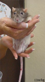
Based on the continuing ulcerations, pain and discomfort for the rat, and the fact that to continue to anesthetize the rat for each removal and redress of the antibiotic wraps posed a risk to her health, the decision was made to amputate both hind legs in order to prevent further deterioration of her overall health.
Outcome
Following the amputation the adult female rat did remarkably well, and was able to get around in her cage without difficulty. She continues to live in good health with her cage mate (who had undergone surgery for removal of a mammary tumor) both of which have been officially adopted by the vet and her staff.
The etiology of this condition was never discovered and is still believed to be an autoimmune disorder, particularly in light of the fact pin point lesions have begun to be noticed on the front limbs of this little rat as of this writing, and her cage mate never showed any similar signs or symptoms of the condition.
Follow-up
This rat has been restarted on prednisone for the pinpoint lesions on the front limbs. When her condition becomes such as to impact her quality of life, euthanasia will be chosen. It has been decided that at that time a necropsy will be done and tissue samples will be sent to pathology to try and determine the cause of the disease process if possible.
Photos
|
 |
 |
||
|
Photos 1 & 2 above show inflammation and excoriation of both hind limbs and feet at the start of treatment. Photo 3: Shows healed amputated hind limbs following nearly 12 weeks of persistent oral and topical treatments. |
||||
Case history and photo permission courtesy of Natalie Rabiner, DVM and Alamo Veterinary Clinic

