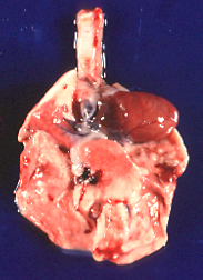
Figure 1: Pathology of Car bacillus
Photos courtesy of IDEXX RADIL
 Photo 1: The bronchopneumonia from CAR bacillus infection is similar to that of mycoplasmosis. The right middle lobe appears to be the most commonly affected site. |
 Photo 2: Peribronchiolar lymphoid hyperplasia with transmucosal lymphocyte migration as in mycoplasmosis is suggestive of CAR bacillus. Silver stains and immunofluorescent techniques will demonstrate the organism (arrow) in tissues. |
Slides and descriptions courtesy of University of Missouri-University of Missouri – Comparative Medicine Program and IDEXX-BioAnalytics (IDEXX RADIL), and Craig Franklin, DVM, PhD, Associate Professor.


