
Figure 5: Mesentery Lymphoma in near 2-year-old female rat (Rosina).
Case history and photos
History
Rosina, a female rat, 20 days short of 2 years old adopted by owner at 4 months old from a reputable breeder in Spain. Rosina gave birth to only one litter at 7 months old and did wonderfully well. Rosina was housed with 7 cage mates that included two of her offspring. Up until the last week of life, she had no previous history of illness requiring treatment other than usual parasite control.
Clinical Signs
The last week of life, owner noted, paleness of ears, a lump felt in the area of the uteri, and some vaginal discharge on the 23rd of June. She was taken promptly to vet.
Diagnosis
Veterinarian assessment: mass palpated, suspicious for tumor, suspicious for pyometra.
Plan: Placed on Bactrim and meloxicam, to be discharged to home, and scheduled for ovariohysterectomy for the following week.
Once home the owner noticed Rosina wasn’t getting any better and was brought back to be admitted on the 30th of June for close monitoring. Surgery was then set for ovariohysterectomy on the 2nd of July.
Treatment
Rosina was given the preop medication midazolam, and surgery performed under isoflurane anesthesia. The intent was of removing the infected uterus; however the reproductive tract was healthy-looking, but a large mesenteric mass was discovered. Video call placed to owner to explain and show findings while rat under anesthesia. A mutual agreement was made to humanely euthanize her without waking her up from surgery. The tumor was collected and sent off to the lab
Outcome
Euthanasia.
Follow Up
A biopsy with histopathology:
Histopathology Report
Clinical History
A rat with a large mass in the mesentery. Enlargement of several lymph nodes.
Clinical suspicion: Lymphoma
Necropsy profile
Mesenteric + Lymph nodes
Diagnosis
Mesentery: large cell lymphoma, high grade
Small Intestine: mild diffuse lymphocytic enteritis
SAMPLE
Excisional.
A block measuring 8.0 x 6.5 x 4.0 cm was received from the stomach and intestine. Within the adipose tissue adherent to the organs, a grayish-brown, lobulated mass measuring 5.0 x 5.0 x 4.5 cm was identified. The cut surface was partially white, solid, and firm, and partially reddish, solid, and softened.
Partial inclusion
Fragments No.: 16; Bock No.: 8.
Cassettes:
A1 to A4 – mass
A5 – intestine
A6 and A7 – largest fragment
A8 – smallest fragment
MICROSCOPIC DESCRIPTION
Eight sections of small intestine and mesenteric fat are present and examined.
The mesenteric adipose tissue is extensively expanded and effaced by a densely cellular, non-encapsulated, infiltrative neoplasm composed of sheets of large lymphoid cells. The neoplastic cells are supported by a scant fibrovascular stroma and exhibit moderate to marked cellular and nuclear pleomorphism. Cells are round to polygonal with distinct cell borders, a moderate amount of lightly eosinophilic cytoplasm, and large round to oval nuclei containing coarsely stippled to vesicular chromatin and one to several prominent nucleoli containing coarsely stippled to vesicular chromatin and one to several prominent nucleoli. Anisocytosis and anisokaryosis are moderate to marked. Mitotic activity is moderate, with up to 3 mitotic figures per high-power field. Numerous individual apoptotic neoplastic cells are present throughout the tumor and are frequently surrounded by tangible body macrophages, imparting a diffuse “starry-sky” appearance. Multifocal to coalescing areas of coagulative necrosis affect approximately 30% of the neoplastic tissue. The adjacent intestinal wall is not infiltrated by neoplastic cells and maintains normal architectural organization. The only intestinal alteration consists of a mild, diffuse lymphocytic infiltrate within the lamina propria, consistent with mild diffuse lymphocytic enteritis.
GENERAL COMMENT
The mesenteric mass is consistent with a high-grade large cell lymphoma arising within the mesenteric adipose tissue. Features supporting a high-grade lymphoma include the large cell morphology, moderate to marked cytologic atypia, frequent apoptosis resulting in a prominent starry-sky appearance, and extensive tumor necrosis. The absence of neoplastic infiltration within the adjacent intestinal wall suggests that the lesion is centered in the mesentery rather than representing secondary extension from an intestinal lymphoma. Immunophenotyping (e.g., CD3 and PAX5/CD79a/CD20 immunohistochemistry) may be performed if confirmation of T- versus B-cell lineage is required.
Pathology report by permission: Vedis Laboratory
Photos

In row 1, the left and right photos show Rosina at a healthy young age. |
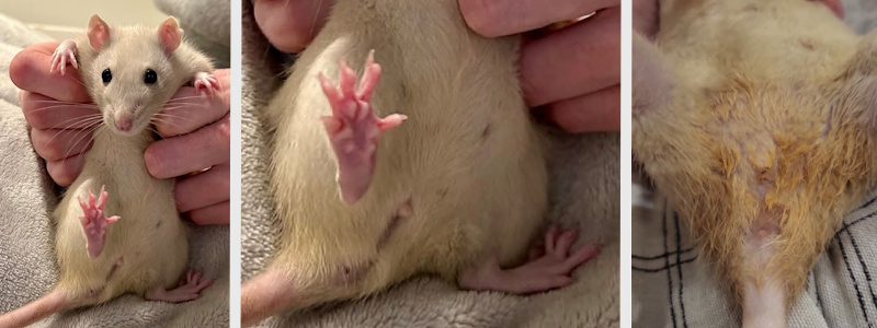
In row 2, the photo on the left (with close-up) was taken prior to any signs of illness. The photo on the right was taken at almost 2 years old and shows discharge, a possible sign of pyometra, upon which the rat was taken to a veterinarian. |
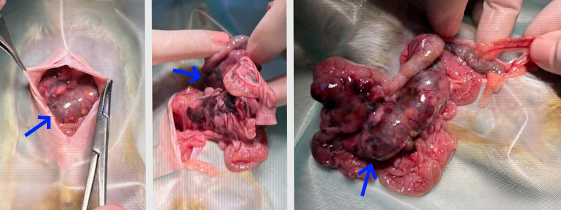
In row 1, the photos taken by the veterinarian during and after (postmortem) what was intended to be an ovariohysterectomy surgery. When the large mesenteric mass (blue arrows) was discovered the rat’s owner was contacted and the decision was made to euthanize the rat without letting her wake up from surgery. Histopathology conducted on the mass found features consistent with a high-grade large cell lymphoma arising within the mesenteric adipose tissue. |
Case courtesy of: Barbara Pinto (owner)
Veterinary Surgeon: Barbara Tomaras DVM
Histopathology report courtesy of: Vedis Laboratory
Photos courtesy of: Barbara Pinto (owner), Barbara Tomaras DVM, and Ipar Argiak Rattery
Photo compilation by Cyzahhe
Case editing courtesy of Karen Grant RN

