Figure 2: Mammary tumor in female rat (Pooka).
Post-op photos of incision site
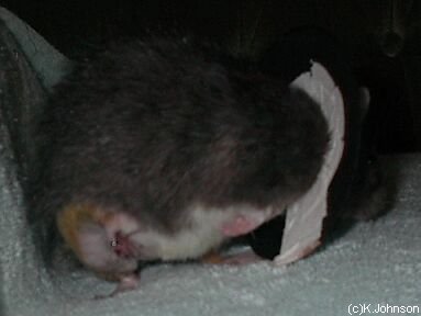
The photos shown here were taken following the removal of a mammary tumor.
Pooka seen here is approximately 18 months old. It is common to see mammary tumors start to develop in this age group. The area of mammary tissue in rats is extensive, and as seen below, shows the location of this tumor removal being to the right and lateral of the abdomen.
Depending on tumor size, and the involvement, a long wide excision may be necessary in removing all of the tumor, since they can grow to be quite large.
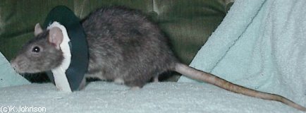
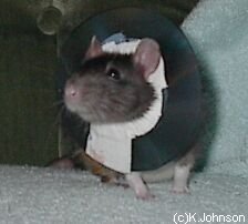
Due to the location site on the body, steel sutures tied individually were chosen over other monofilament or multifilament type suture material, and an Elizabethan collar applied to further prevent this rat from chewing or licking the incisional area. It is generally not recommended in most cases to use an Elizabethan collar in rats since it prevents them from holding their food, and inhibits the intake of nourishment.
Photos
 Photo 1: Incision with clip showing seen right and lateral of abdomen. |
 Photo 2: Additional view of incision showing clips. |
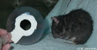
 Photo 3: Showing side view of Elizabethan collar. X-ray film paper can be cut to size and kerlex or tape applied around edge that will encircle the neck. |
 Photo 4: Front view of Elizabethan collar. |
 Photo 5: Pooka managed to remove her collar herself as soon as she arrived home. |
Case history and photo courtesy of Kristin J. Johnson The Wererat’s Lair


