Megacolon Figure 3b

Figure 3b: Necropsy of a 19-month-old rat with megacolon

pre-necropsy

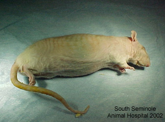
Photo 1: Shows this rat 24 hours following expiration.

incision

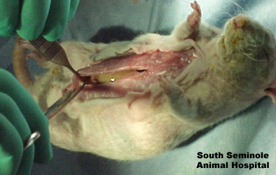
Photo 2: Shows initial midline incison.

internal view

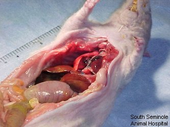
Photo 3: Shows internal organs.

enlarged colon

Photo 4: Shows distended colon.

colon

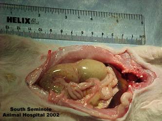
Photo 5: Shows another view of distended colon.


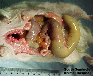
Photo 6: Shows gross distention of colon.

ruptured colon

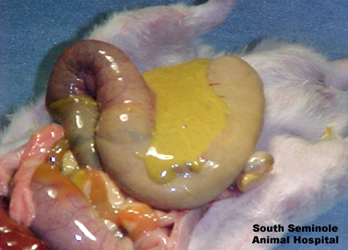
Photo 7: Shows gross rupture of colon and contents.

diagram of organs

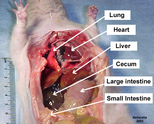
Photo 8: Shows diagram of organ placement in the thoracic-abdominal cavity.

Photos courtesy of Bellaratta’s Nest Rattery and Dr.David Byrd DVM, South Seminole Animal Hospital

Cross-references

Linked from

Disclaimer

The Rat Guide and its affiliates accept no responsibility for misuse or misunderstanding of its information. This guide in whole or part, exists solely for the purpose of recognizing and understanding the care and illnesses in the pet rat. Please seek advice and treatment from a qualified Veterinarian if your rat is ill.

2000 - 2024 by Karen Grant RN. All rights reserved.
All other written and visual materials used by permission of specific authors for the sole use of the Rat Guide. Please visit our Privacy Policy for details.
Brought to you by KuddlyKorner4u
See Logos page for linking to the Rat Guide.
Contact us here: Rat Guide Team
Please note: Rat Guide email is not checked daily. Send e-mail to if you have an urgent medical problem with your pet rat. When possible, it is always best to take your rat to a qualified rat veterinarian.