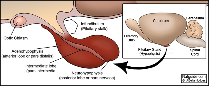
Figure 1: Anatomy of the rat’s pituitary gland and diagrams.
|
|
| Diagram 1: Indicates the different lobes of the pituitary gland. | Inset: Shows the brain and location of the pituitary gland. |
The pituitary gland in rats, as in other mammals, is situated in a bony cavity called the sella turcica. However, the diaphragma sellae (a horizontal fold of dura mater) which covers and acts as a roof of the sella turcica retaining the pituitary gland, in other mammals, is normally an incomplete structure in the rat.
In rats, as in other mammals, the pituitary gland is composed of the adenohypophysis and neurohypophysis. The adenohypophyis is represented by the anterior lobe (pars distalis), the intermediate lobe (pars intermedia) which is only rudimentary in humans, and the infundibulo-tuberal lobe (pars tuberalis). The neurohypophysis is composed of the posterior lobe (pars nervosa), the median eminence, and the hypophyseal stalk.
Control of the pituitary gland is under the influence of the hypothalamus. The secretion of hormones by the anterior lobe of the pituitary is regulated by releasing hormones from the hypothalamus via the hypophyseal portal veins. Because there is no such portal vein connection in the neurohypophysis control by the hypothalamus is through nerve impulses.
Each lobe of the pituitary produces and secretes hormones that regulate many of the body’s functions, as well as to stimulate other endocrine glands. The hormones produced by the pituitary gland are not all secreted in continuous fashion. Many of the hormones are released in bursts, alternating between periods of activity and inactivity (Merck, 1998).
Staining techniques reflects three cell types in the anterior lobe categorized as: acidophils, basophils, and chromophobes. The addition of newer immunostaining techniques shows the specific hormones contained in those cell types, and in the lobes of the pituitary gland:
Anterior lobe (adenohypophysis or pars distalis)
- Acidophils (cells containing glycoprotein hormone):
- Somatotrophs (the predominant cell type in the anterior lobe of the male rat) (Mohr, et al., 1992)
- GH cells: produce growth hormone.
- Lactotrophs (more numerous in the female rat) (Mohr, et al., 1992)
- PRL cells: Produce prolactin which stimulates the corpus luteum (temporary endocrine structure in the female reproductive system) to produce progesterone and stimulate the production of milk in the mammary glands.
- Somatotrophs (the predominant cell type in the anterior lobe of the male rat) (Mohr, et al., 1992)
- Basophils (cells containing glycoprotein hormones):
- Gonadotrophs:
- LH: Lutenizing Hormone which in females is associated with the maturation of the follicles, the manifestation of heat (or estrus), and the release of the egg. In males, the lutenizing hormone stimulates the testes to release testosterone.
- FSH: Follicle-Stimulating Hormone that stimulates estrogen secretion in females and spermatogenesis in males)
- Thyrotropin or TSH: thyroid stimulating hormone stimulates thyroid gland to release thyroxin (T4). Thyroxin regulates the metabolism.
- Corticotropin or ACTH (adrenocorticotropic hormone which stimulates adrenocortical growth and its production of glucocorticoids, mineralocorticoids, and androgens).
- Gonadotrophs:
- Chromophobes:
- Cells believed to already have released hormones.
Intermediate lobe (pars intermedia)
- MSH (melanocyte stimulating hormone which influences formation and deposit of pigmentation).
- Beta-endorphinendorphin (“endogenous morphine”): one of four groups of endorphins (alpha, beta, gamma and sigma). Beta-endorphins are found in the pituitary gland. It is a most potent peptide opioid that acts as a neurotransmitter affecting both central and peripheral nervous systems to reduce the sensation of pain. It is thought to affect appetite, release of sex hormones, and stimulate the immune system.
Posterior lobe (neurohypophysis or pars nervosa)
- Oxytocin (stimulates contraction of smooth muscle in the uterus and ejection of milk from the mammary glands).
- Arginine Vasopressin or anti-diuretic hormone (stimulates smooth muscle of blood vessels and digestive tract and promotes water retention by way of the renal tubules maintaining water balance and increases blood pressure).
References
- The Pituitary Gland: Introduction. (n.d.). Retrieved December 25, 2008, from http://www.merckvetmanual.com/mvm/index.jsp?cfile=htm/bc/40500.htm.

