Figure 2: Squamous cell carcinoma at base of the ear involving Zymbal’s gland (Ramekin).
Case history and photos
History
Ramekin, a 22-month-old male rat, presented with a large lump on his face just below the right ear.
Clinical Signs
The lump appeared virtually overnight, growing to a diameter of around 1/2 inch. It was believed, at that time, to be an abscess. His general condition was otherwise healthy.
Initial Exam
Physical examination by the vet revealed a very hard lump that didn’t move freely. Upon X-ray, no bone involvement or other features were apparent. Needle aspiration was performed and serosanguinous fluid obtained showing mostly red blood cells, macrophages (cells that have the ability to ingest and destroy particulate substances, such as bacteria) and some seg neutrophils (segmented white blood cell).
Diagnosis
Possible cyst.
Possible tumor formation.
Treatment
Treatment was initially begun by having the cyst drained, and starting a course of enrofloxacin (Baytril), and Cortisone (dexamethasone).
A few days following the initial treatment, the cyst again returned, and drainage was repeated.
DSMO was prescribed, and applied daily to the area of cyst removal. The cyst reappeared after two days.
Follow-up
At this time a recurrent hydrocyst due to trauma to the face, or Zymbal’s gland tumor possibly squamous cell in nature was suspected and the decision made to do surgery.
Exploratory surgery was performed:
“The cyst explored, was invasive of muscle, nerves, and blood vessels around the ear base. Invaded ear brought up pus and blood. The area was debrided and flushed. As much of the tissue was curretted as possible. A drain inserted in place, too leave for 5 days if possible.”
Outcome
The cyst returned within 3 days post op. It was now filling with fluid rapidly and drainage could be seen coming from the ear canal and incision drain. The tumor itself was visibly growing larger and rapidly below the cyst.
Daily draining of the cyst was performed for several days and during this time Ramekin continued to be otherwise active and eating.
One week after surgery the tumor/cyst hemorrhaged profuse bright red blood from the ear canal, the surgical incision, and the opening made for the drain that had been inserted in the cyst. The decision was made that Ramekin not be allowed to suffer, and was euthanized the following day.
Photos
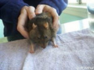 Photo 1: Depicts developing growth noticed here on side of face. |
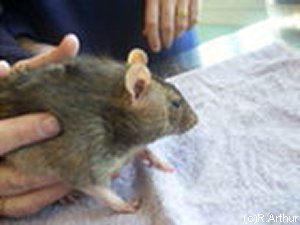 Photo 2: Closer camera view of growth. |
 Photo 3: Tumor development can be clearly seen using the ear and eye as a marker. |
Photos courtesy of Al and Robyn Arthur, of The Dapper Rat, http://www.dapper.com.au
Collaborated case study by, Dr Larissa Ladyko B.V.Sc, and Robyn Arthur.
Diagram drawing of Zymbal’s gland location in rats https://reni.item.fraunhofer.de/reni/trimming/manus.php?mno=034.

