Figure 1: Squamous cell carcinoma of the jaw in male rat (Bob).
Case history and photos
History
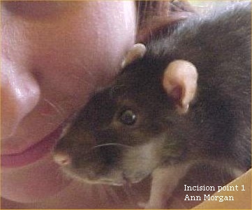
The following photos of “Bob” show quick growth progression of the actual tumor. Despite the graphic nature of these photos it is important to note that “Bob” was not in pain, and was well cared for by his owners.
Clinical Signs
Bob was first noted to have a small lump in April 2002, and was taken to the vet.
Diagnosis
Possible abscess or tumor formation.
Treatment
At this time the Vet excised and drained the growth, believing it to be an abscess produced by Actinobacillis, since on excision white powdery to white fibrous appearing strands were noted. The wound, though, healing slowly, still drained a small amount of serous exudate.
Growth continued and a second trip was made to the vet where another partial excision and suturing was done.
Outcome
A month later in May, a foul putrid odor was noted from the wound. The site was cleaned and the following day the lump that was present appeared to have collapsed and was bleeding. A trip was made to the vet, where tissue for histology was sent. The results returned showing the lump to be positive for squamous cell ca, seen in the photos: 3 and 4 below.
Prognosis given by the vet based on the results and the aggressive growth of the tumor was that “Bob” might only survive for another two weeks. Progression of this type of tumor took only one month.
Photos

|
|
|||
| Photos: 1 and 2 show a large growth on the jaw. | ||||
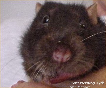 Photo: 3 Frontal view |
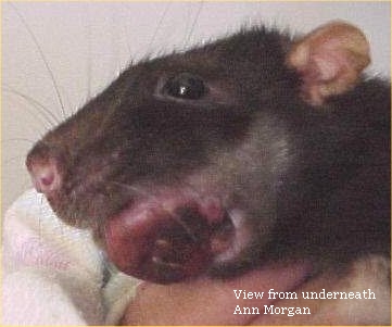 Photo: 4 Viewed from below |
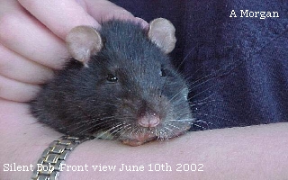 Photo: 5 Swelling can be observed on side of face. |
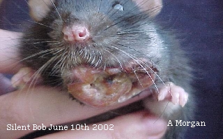 Photo: 6 A full face view |
*note: Bob went to the Rainbow Bridge the second week of June, 2002. Rest sweetly Bob, and thank-you to Ann and Emma for so kindly sharing these photos. Bob was dearly loved by Ann and Emma.
Photos courtesy of Ann Morgan, of Rock-a-Bye Ratties Rockhampton, Australia; http://rockabyeratties.com/
AusRFS reg breeder 017/001


