Figure 5: Fibrosarcoma of the skin in male rat (Sambuca).
Case history and photos
History
Sambuca, a 27-month-old Black blazed (husky-marked) intact male rat. Housed together with another male in a cage setting. Fed my own recipe of rat mix with Oxbow as a base. Pet only, due to megacolon in one sibling. Always a large rat, he became overweight at about eight months of age.
Sambuca has a history of respiratory disease, requiring frequent treatment with antibiotics. He was on Zithromax and doxycycline for three weeks prior to surgery (postponed surgery by two weeks for respiratory symptoms).
Sambuca began showing kidney disease symptoms 10/03: he has increased water intake, increased urine output, and sunken haunches. His diet is supplemented three times a week with bananas and liquid B-vitamins. He also grew a tumor (presumably mammary) just to the right of his midsection.
Clinical signs
Growth on right shoulder region. Tumor first appeared like an abscess, but lancing produced excessive bloody fluid. The surface grew and the tumor took on the appearance of a raised, but flat shiny abrasion. It grew to approximately three centimeters and remained static for one month. He began scratching it frequently, despite nail trims, and caused it to ooze bloody fluid.
Diagnosis
Histopatholgy performed by Michael Garner, DVM, Dipl ACVP of Northwest Zoopath. Diagnosis was “intermediate grade fibrosarcoma, skin”. Microscopic findings described interlacing streams of haphazardly arranged sheets of cells in a collagenous stroma. “Cells had scant eosinophilic cytoplasma and ovoid vesicular nuclei with one or two prominent nucleoli and occasional mitotic figures”. The tumor abutted the deep surgical margin and was focally ulcerated.
Treatment
Surgical removal, 10/15/03. Weight 795g. Pre-medicated with buprinex (analgesic) and glycopyrrolate (anticholinergic) subcutaneously approximately 45 minutes prior to induction. Induced on isoflurane and O2 in a tank, then masked. Sambuca was shaved and the surgical area scrubbed with dilute chlorhexiderm. Surgery was performed on a microwaved heat pad covered with a clean towel. A surgical drape was placed and Ember Couture, D.V.M. performed the surgery with a mask, hat, and sterile apron, gloves, and instruments. Tumor was excised and skin was closed with 5-0 monocryl and staples. Sambuca recovered on oxygen and a heat pad. He was then moved to a heated incubator for recovery. He remained on Zithromax for 21 days post-surgery. Please see step-by-step photos.
Outcome
Fibrosarcomas are almost impossible to completely excise, so it is expected that the tumor will grow slowly. Noted apparent regrowth 12/25/03. Sambuca scratched the area, opening the lump on 12/30/03. The lump was oozing green pus (abscess). Cleaned and flushed the abscess and began oral antibiotic treatment with TMS (trimethoprim-sulfa). Flushed the abscess three times daily (after debriding opening) and packed triple antibiotic ointment subcutaneously overnight for two nights. Once the abscess cleared, it was apparent that the fibrosarcoma was still present, though quite small.
Followup
Sambuca passed on at the age of 2 years, 7 months on 2/10/04. His fibrosarcoma had grown to about the size of a small marble. His midsection tumor continued to grow, but it did not appear to cause him any discomfort.
Photos
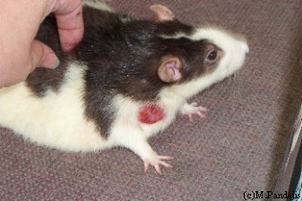 Photo 1: One week pre-op. |
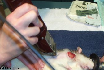 Photo 2: Prepping for surgery. Sambuca gets a shave under Iso (isoflurane gas). |
 Photo 3: Sambuca shaved and unconscious. |
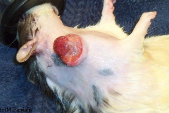 Photo 4: Sambuca scrubbed and ready for surgery. |
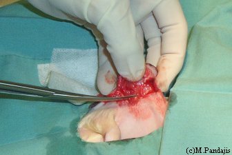 Photo 5: Draped, skin open, Dr. Couture begins excision. |
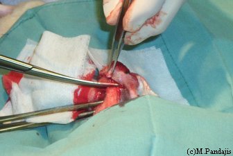 Photo 6: Sambuca’s tumor excision progresses. Note clamps on major vessels. |
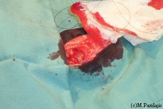 Photo 7: Tumor removed, Dr. Couture begins subcutaneous closure (dissolvable stitches). |
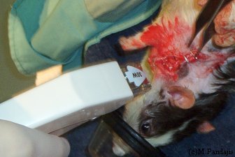 Photo 8: Dr. Couture pinches outer skin together for proper stapling to close. |
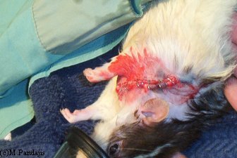 Photo 9: Sambuca fully stapled and ready to switch to oxygen. |
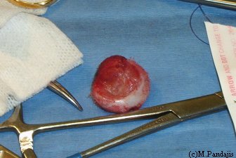 Photo 10: The excised tumor. Approximate width is 3cm. |
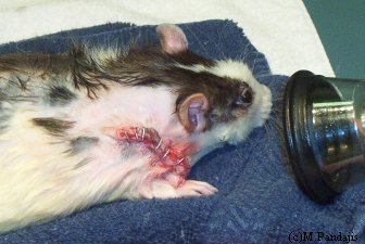 Photo 11: Sambuca recovers with the help of oxygen. |
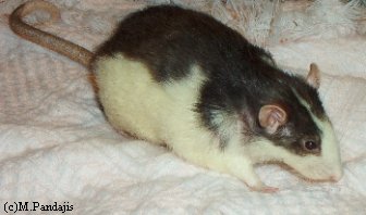 Photo 12: Sambuca fully healed. |
Case history and photos courtesy of Melissa Pandajis
Acknowledgments and thanks to the following veterinarians for their contribution:
Ember Couture, DVM, Cats and Critters, Rochester, NY
Michael Garner, DVM, Dipl. ACVP, Northwest Zoopath, Monroe, WA

