Figure 2: Bladder stones
Case history and necropsy photos
|
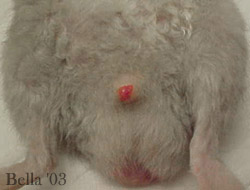
Photo 1: Blood oozing from the penis.
|
|
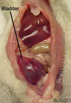
Photo 2: The bladder in its natural position. Even before removal its size and characteristics are obviously abnormal. |
|
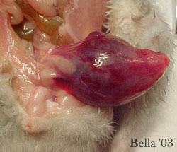
Photo 3: The bladder is pulled away from the body for examination. It is 4-6 times normal size. Keep in mind this is after the removal of 6 mL of fluid.
|
|
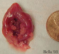
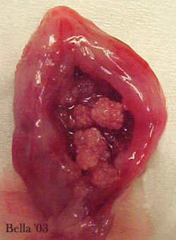
Photo 4: The bladder removed and opened. Note the severe inflammation of the entire bladder and the crystals within it.
|
|
Photo 5: In this close up you can see the abnormal thickening of the bladder wall, which are usually thin and pliable. The bladder is completely packed with crystals.
|
|
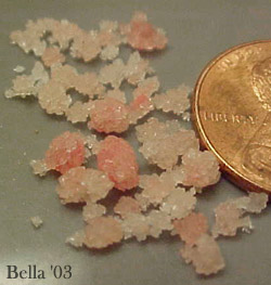
Photo 6: A close up of struvite crystals after removal from the bladder.
|

|
Photo 7: A view of a normal full bladder from an adult male rat (held with gloved fingers). Note the thin balloon-type structure of the bladder and its transparency.
|
Emergency veterinarian- Dr. Frank Williams, DVM/FW (Emerald City Emergency Clinic – Seattle Washington)
Consultant veterinarian- Dr. Richard Mckinniss, DVM (South Seminole Animal Hospital – Fern Park, Florida)
Case history and necropsy: Joanne “Bella” Hodges

