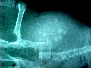
Figure 1: Bladder stones in 8-10 week old male rat (Samuel).
Case history and X-rays
History
Samuel, a juvenile genetically tailless, blue rex male was air shipped from Northern California to Seattle Washington with 5 other rats. His age was approximately 8-10 weeks old. He had no access to free fluids during the time in the shipping container (estimated 10-12 hours). Apple slices and carrots were supplied during containment.
Clinical Signs
After he was picked up at the airport it was noticed that he had hematuria (blood in the urine). Samuel was given a dose of Baytril. He was also treated for lice (topically) along with the rest of the rats. Samuel was given oral fluids in an attempt to hydrate him.
Several hours later, after being moved into a cage with 2 other rats, frequent squeaking was heard as if the rats were squabbling. It was then noticed that Samuel was squeaking and licking himself with no other rats in the immediate area.
Samuel was removed from the cage. There was no evidence of urination. His bladder felt hard and enlarged indicative of a possible urinary blockage. Samuel stayed on his back when held and only moved when waves of pain gripped him. As the pain came he would ball up, frantically lick at his penis, and bite his paws and feet. Samuel was given infant Tylenol to take the edge off the pain and was transported immediately to the emergency vet clinic.
Initial Exam
Physical examination by the veterinarian revealed a very hard abdominal mass. The vet suspected urethral blockage. Possible suspected causes were:
- UTI (urinary tract infection)
- uroliths
- neoplasia (tumors)
The X-rays showed a highly distended bladder filled with uroliths. The emergency vet noted in his case notes that “this problem obviously did not happen overnight.”
A 25-gauge butterfly catheter was used to remove 6 mL of urine via cystocentesis. (This can be a risky procedure with an extremely distended bladder. There is a chance that the needle could lacerate the tissue causing urine to escape into the abdominal area.)
Urinalysis results:
- Color: brown
- Character: turbid +++
- Specific gravity: 1.018
- pH: 8.0
- Protein: +++Blood: Large
- WBC: 2-6/hpf
- RBC: TNTC
- Crystals: struvite crystals 6-10/hpf
Diagnosis
- Struvite crystal formations in the bladder
- Urethral obstruction
- Possible chronic UTI
Treatment
Possible treatment would be immediate surgery to remove the crystals from the bladder and clear the blockage.
Long term prognosis included a strong possibility of the crystals returning.
Outcome
We elected to have Samuel euthanized.
Samuel’s necropsy showed a severely inflamed and distended bladder completely filled with sharp struvite crystals.
The breeder who shipped Samuel has decided to cease the shipping of rats.
- Necropsy photos (graphic). See Figure 2
Case Review
Dr. Mckinniss DVM evaluated the report, X-rays, and necropsy photos. He concluded that, due to the young age of the rat and the severity of the condition, that there was most likely a congenital (possibly genetic) cause for Samuel’s condition.
It is speculated that the final blockage was brought on by the lack of fluids during shipping and containment.
 In this X-ray you can see that the bladder is 4-5 times larger than normal. |
 Note the density of the struvite crystals. Being one dimensional, this view does not even come close to showing the actual amount of crystals harbored in the bladder. |
Emergency veterinarian- Dr. Frank Williams, DVM/FW (Emerald City Emergency Clinic – Seattle Washington)
Consultant veterinarian- Dr. Richard Mckinniss, DVM (South Seminole Animal Hospital – Fern Park, Florida)
Case history and necropsy: Joanne “Bella” Hodges
Thanks go to Annie Braun for financing Samuel’s emergency care and to the NW Ratspack for their assistance in this case.


