Figure 1: Zymbal’s gland tumor in 18-month-old male rat (Joey).
Case history and photos
History
An 18-month-old, 583 g, Russian blue Burmese Dumbo male rat with no known illnesses.
Clinical Signs
Facial swelling on left cheek eventually becoming a large mass exhibiting ulceration and necrosis. The rat only exhibited overt signs of pain during wound care and in the last few days of life.
Time from onset to euthanasia: 4 weeks.
Diagnosis
Probable Zymbals’ gland tumor.
Treatment
After the initial swelling was noticed, an incision was made and pus was drained. The wound was irrigated daily with diluted Betadine and the rat was put on antibiotics (Baytril 10 mg/lb and Clavamox 6.5 mg/lb). Banamine and eventually buprenorphine were administered for pain management.
Outcome
Due to Joey’s deterioration in condition, and increasing pain, euthanasia was elected by owner.
Follow-up
NECROPSY
Large facial mass. Probable Zymbal’s gland tumor. Caudal liver lobe: orange and mottled.
HISTOPATHOLOGY SUMMARY
Zymbal’s gland carcinoma
Zymbal’s gland:
Carcinoma of the Zymbal’s gland characterized by circumscribed mass of irregular acini or solid sheets of cells containing cystic cavities filled with sebum, keratin and necrotic cells; neoplastic sebaceous and squamous cells are often pleomorphic and anaplastic; multifocal areas of papillary projections of squamous epithelium in cystic spaces; multifocal areas of branching cords of cells showing squamous differentiation; multifocal areas of acini consisting of solid sheets of fusiform cells with elongated nuclei and scant cytoplasm; focal regions consisting of predominantly anaplastic spindle cells with small foci of neoplastic squamous epithelium; multiple regions showing transition from neoplastic squamous epithelium to anaplastic, pleomorphic cells with occasional mitotic figure; multifocal regions of inflammatory eosinophils and lymphocytes in the well formed stromal cells.
Photos
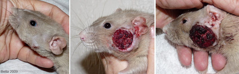 Progression of cancerous mass involving Zymbal’s gland. |
Histopathology: by IDEXX RADIL
Necropsy, and photos: Joanne “Bella” Hodges

