Figure 1: Glomerulonephritis in 20-month-old male rat (Martin).
Case history and photos
History
A 20-month-old, 340 g, American blue Dumbo intact male with no previous health history.
Clinical Signs
Over a 4-month period: pale extremities, steady gradual weight loss, lowered body temperature, and lethargy. His appetite was good, until the last few weeks, and he still sought out human interaction.
End Stage (last few days)- Decrease in appetite, listing to one side, left eye partially closed, rapid weight loss, lightened color on incisors.
Diagnostics
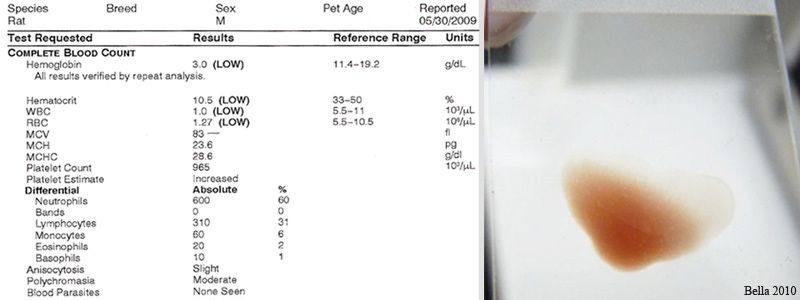
Blood was lactescent (milky). The packed cell volume (PCV) was very low, blood work showed lipemia and anemia.
Treatment
Prophylactic antibiotics, dexamethasone, and an iron-enriched vitamin supplement.
Outcome
Martin did not respond to treatment and the owner opted for euthanasia.
Follow-up
NECROPSY:
Blood and bone marrow were collected pre-mortem. Euthanasia was pentobarbital IP with the full necropsy performed immediately.
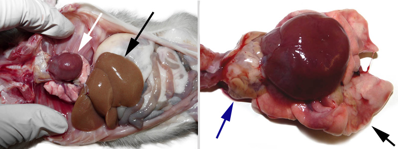
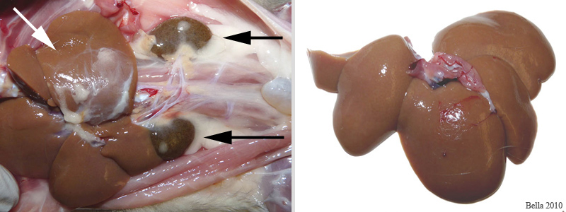
Heart was enlarged and misshapen, liver was pale, pancreatic tissue was yellow, kidneys were a pale brown- when bisected the cortex was light brown/yellow and the medulla was pale. The colon and cecum had black content. The cecum had several lesions.
HISTOPATHOLOGY SUMMARY:
Marked glomerulonephritis was found. Anemia can result from kidney failure due to the lack of erythropoietin production by the peritubular capillary endothelial cells. Anemia can produce the appearance of pale organs and cardiomyopathy can also result from chronic anemia as the cardiac myocytes enlarge in an attempt to pump more blood to oxygen starved tissues, resulting in increased blood pressure which increases the rate of glomerular damage. The cycle, although done by the body to relieve the lack of oxygen from anemia, hastens the damage to the kidneys.
Kidney:
There is marked glomerulonephritis of the kidney. Almost all glomeruli are greatly enlarged and fluffy appearing with the mesangium and basement membranes being thickened with a pale eosinophilic material. There is loss of celluarity and many glomeruli have adhesions between Bowman’s capsule and the surrounding tissue. Bowman’s space is enlarged in many glomeruli. There is widespread loss of renal tubules but remaining tubules have markedly thickened basement membranes, dilated, and contain eosinophilic proteinaceous casts. Interstitial fibrosis and interstitial lymphoid infiltrates are prominent and multifocal areas of mineralization can be seen.
Liver:
Occasional pigment-laden macrophages are within the connective tissue of the capsule.
Photos
 Pallor of the ears, nose, extremities & scrotum can be seen due to decrease in oxygen as a result of anemia. |
 Lab results of Martin’s blood work. To the right is a photo of a drop of blood used to make a slide showing the milky color, thinness, and the oily quality of the blood. |
 First photo: The white arrow shows the enlarged heart and the black arrow shows the liver . Second photo: The blue arrow is pointing to an enlarged thoracic lymph node and the black arrow is pointing to a lung lobe. In this photo the enlarged heart is quite obvious. |
 First photo:: The white arrow is on the liver and the black arrows are on the kidneys. Second photo: Showing the liver after it has been removed. Both of these organs, in a healthy rat, should be a deep, dark red. |
 The above photos are of both of Martin’s kidneys bisected showing the extreme damage. |
Owner: D. Kohn
Vet consultant: Dr. R. Mackinnis
Histopathology: IDEXX RADIL
Necropsy, photos, and micrographs by Joanne “Bella” Hodges


