Figure 2h: Mandible abscess stemming from probable tooth root infection in female rat (Opossum).
Case history and photos
History
“Opossum” was still fairly young when she was adopted from a pet store in early 2009. She was up for “adoption” rather than sale due to chronic eye issues. Although her eye was never normal, it did not require treatment after adoption.
Opossum was fairly “stand-offish”, so her keeper was not aware that something was wrong until she noticed a large scab on Opossum’s right lower jaw in November 2011. The scab was soaked off/removed and the area was flushed with dilute chlorhexidine twice a day. Opossum was put on Baytril.
Clinical Signs
Six days after discovery of the scab – although the abscess area was closing up – an area of necrotic bone had developed.
Diagnosis
Mandible abscess stemming from probable tooth root infection.
Treatment
After consulting another veterinarian Opossum was switched to gentamicin, as it has better bone penetration.
A week later the decision was made to remove the right lower incisor and debride the necrotic bone. Anaesthesia was maintained by administering isoflurane through a make-shift nose cone (thus allowing access to the mouth). Dental elevators were used to remove the incisor. The necrotic portion of the mandible was actually slightly displaced. Dental forceps were used to debride it. The area was left open to allow continued flushing.
Opossum was placed on oxygen during the recovery period.
Outcome
Opossum seemed more comfortable after the surgery. Although her upper incisors were very short and blunt, she did well eating mushy foods. The wound area closed up somewhat, but never healed completely. Opossum also had respiratory issues and was maintained on gentamicin injectable, with subQ fluid injections to help support her kidneys.
Opossum passed away in January 2012.
Photos
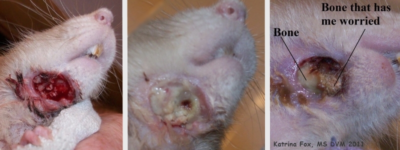 The first photograph was taken after the scab was loosened by soaking and removed. The second photograph was taken the following day. The third photograph was taken six days after discovery and removal of the scab (and twice a day flushing); it shows the abscess area closing up, as well as the area of necrotic bone that is of concern. |
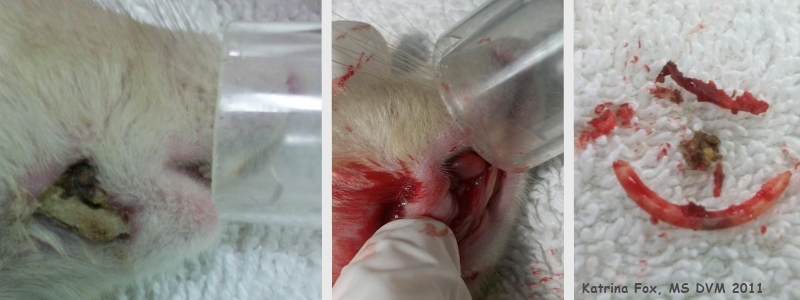 The two photograph on the left show the make-shift nose cone that was used to maintain anesthesia. The first photograph was taken pre-op and shows the displaced necrotic portion of the mandible. The second photograph was taken during the surgery. The photograph on the right shows the incisor and bone that were removed. |
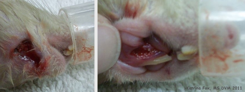 These two photographs were taken post-op, during recovery. Note that the upper incisors are very short and blunt. |
Case history and photos courtesy of: Katrina Fox, MS DVM
Case Editors: Karen Grant RN, and Cyzahhe

