Figure 1: Auricular (ear) polyp in male rat (Lukkas).
Case history and photos
History
Lukkas had been healthy up until the age of approx. 1 1/2 years old. At that time his cage mate died and Lukkas almost died of what we suspect was mycoplasma related pneumonia. He recovered from this initial illness, but relapsed several months later. These two bouts of pneumonia left him quite frail.
Clinical Signs
Lukkas was noticed to have a “foul” odor with pus coming from the left ear. He did not have any head tilt.
Diagnosis
Initial examination by the vet diagnosed an ear infection. Subsequent diagnosis auricular polyp.
Treatment and Follow-up
Lucas was prescribed oral antibiotics (Baytril 5 mg. BID) and Tresederm drops (3 in the afflicted ear daily).
After approx. 1 week of treatment the foul odor was almost completely gone as well as the pus. Upon examination one day, I found a growth protruding from the ear canal. It was dark red and rounded with the surface broken and a scabby area at the tip where he had been scratching at it.
Lucas returned to the vet where the growth was partially removed and diagnosed, at this time, as a nasopharyngeal polyp. There was minimal bleeding at the time of the removal. The polyp most likely formed in the eustachian tube.
Outcome and Follow-up notes
The problem noted with partial removal was if the base (source) of the polyp isn’t removed then 50% to 75% of the polyps will reoccur in 1 to 8 months. A more complicated, but thorough, surgical procedure called a ventral bulla osteotomy is recommended in larger animals with this condition. This surgery removes the lower part of the bone that surrounds the inner ear. However in a rat this would not be feasible since the bones are so minute.
Lukkas continued on the Baytril, and taken off the Tresederm drops. He was prescribed Panalog ointment twice a day in the affected ear.
As predicted the polyp did grow back within several weeks. The foul odor never completely went away. Lukkas’ respiratory condition worsened. He began to have trouble walking, and bleeding was noted coming from his ear. Doxycycline was added at this point to his treatment. Lukkas did have a partial recovery, but then relapsed again. It was at this time that the difficult decision was made to euthanize Lukkas to prevent his further suffering.
Photos
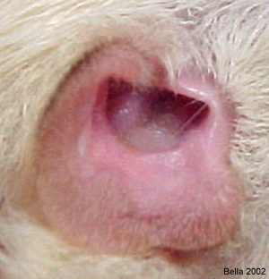 Photo 1: Pus and wax can be seen just inside left ear canal |
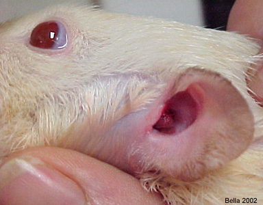 Photo 2: Note pink protrusion from left ear canal. |
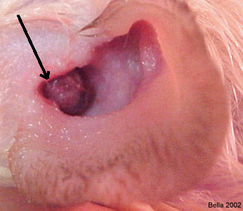 Photo 3: Protrusion more pronounced. |
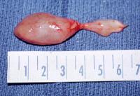 Photo 4: This photo, courtesy of John E McClay, MD, Assistant Professor, Department of Otolaryngology, Division of Pediatric Otolaryngology, Children’s Medical Center, University of Texas at Southwestern, shows an intact nasopharyngeal polyp removed from a pediatric patient. The large bulb mass to the left is the body of the polyp and as you view towards the right of the photo, shows stem and base. |
|
Photo 5: Shows polyp size recreation in rat. |
Case history and photos courtesy of J. “Bella” Hodges, Bellaratta’s Nest Rattery
Pediatric polyp photo and study can be found at https://emedicine.medscape.com/article/994274-overview

