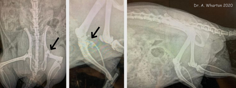
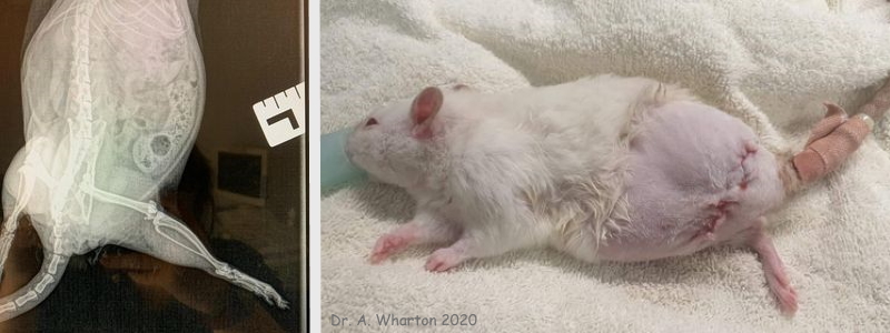
Figure 1: Amputation in 1-year-old female rat (Peaches)
Case history, photos and video
History
Peaches is a 1-year-old female rat owned by Emma Andrews. Her original injury is unknown.
Diagnosis
Peaches was brought in to Dr. Charlie Lyons with an acutely lame left hindleg that appeared to be rotated outwards. Peaches was still trying to use the limb, although it appeared to be very painful. Dr. Lyons could feel the hip luxating, which was confirmed on x-ray. She also identified abnormalities in the stifle (the equivalent of the human knee joint) on the same leg.
The x-rays were shared with Dr. Adele Wharton. Both veterinarians agreed that amputation would be the best option, since the stifle abnormalities meant that femoral head and neck excision would be unlikely to give Peaches a functional limb.
Treatment
The amputation surgery was carried out by Dr. Wharton. The dislocation made it very challenging to locate and elevate the femoral head, but once this was done it was possible to preserve the majority of the thigh muscle to achieve a good cosmetic outcome, as well as a functional one for Peaches. Peaches was administered buprenorphine pre-op, anesthesia was induced and maintained with iso/O2, she received IV fluids throughout the surgery, as well as meloxicam during the surgery and back at home. Muscle closure was with 4/0 PDS, skin and vessels 4/0 vicryl.
Outcome
Recovery was uneventful and, although Peaches was initially a little unsteady on her three legs, she appeared to be much more comfortable.
Follow-up
Dr. Wharton and Peaches’ keeper kept in touch after the surgery for check-ups. Less than 48 hours after the surgery Peaches was already running around again like nothing had happened. At her 72 hour post-op check she was healing nicely, very active, and showing no signs of pain.
Photos and Video

Row 01: These x-rays were taken to diagnose Peaches’ injury. They show the dislocated left hip (first arrow) and abnormal stifle (second arrow). |

Row 02: The first photo is another x-ray showing the dislocated left hip. The second photo shows Peaches immediately after her amputation surgery; she was only on oxygen at that point awaiting recovery. The elastoplast on her tail is holding her IV cannula in place (it was left in until she was taken home). |
This video, taken 48 hours after Peaches’ amputation surgery, shows her running around like nothing has happened. She does not appear to be in pain.
—
Case history, photos and video courtesy of:
Adele Wharton BVSc, MRCVS, CertGP (F&L) Veterinary Surgeon
Charlie Lyons BVetMed, MRCVS
Owner: Emma Andrews
Case and photo compilation by Cyzahhe
Case editing courtesy of Karen Grant RN and Cyzahhe


