Figure 1: Secondary glaucoma in male rat (Gogol).
Case history and photo
History
Gogol is a 3-month-old male rat.
Clinical Signs
Gogol’s right eye started appearing slightly larger than the left in early April. The condition slowly escalated over the next month.
The left eye was much darker (more brown in appearance) while the right eye had a bright red color. The iris appeared slightly yellow compared to that of the left.
Veterinary treatment was not sought at this point because Gogol did not show signs of pain or discomfort, and could see well out of the eye.
May 7
The eye became cloudy.
May 9
Gogol was taken to a vet where the following tests were performed:
- Schirmer tear test (to test for dry-eye)
- Blood test (to rule out diabetes)
- Fluorescent eye stain (to check for ulcers on the cornea)
- Tonometry (pressure test to check for glaucoma)
The eye stain showed irregularity in the shape of the eye which had healed over with an outer skin membrane and was therefore no longer an ulcer. It is suspected that there had been an ulcer at some point.
The tonometry showed the pressure of Gogol’s left eye to be 25 and right eye to be 37 (Normal pressure in a rat’s eye should be between 21 and 26).
Diagnoses
Secondary glaucoma.
The presence of a healed eye injury and glaucoma suggest that there was a trauma to the eye at some point, possibly from pressure being applied at birth or from play or a fall. The injury healed over but left the shape of the eye compromised. The irregular shape allowed pressure to build and glaucoma to form.
Treatment
Trithalmic/Cortisone Opthalmic Ointment (to be applied to both eyes once daily).
Pilocarpine Opthalmic Solution 1% to be applied to the right eye only twice daily.
Outcome
Due to the tests, a small pool of blood formed behind the cornea. However, this drained after three days. The eye decreased in size, gained a more almond-like shape (as opposed to the strong circle), the cloudiness dissipated, and Gogol regained the ability to close the eye completely.
On 13 May, a follow-up was done by the vet to check for any adverse reactions to the medication, such as a recurring ulcer, and another tonometry was preformed. No ulcers were present, and the new pressure readouts were 21 for the left eye and 26 for the right.
Use of the ointment was discontinued and application of the Pilocarpine drops continued for another week before being reduced to once daily and then also being discontinued.
Photos
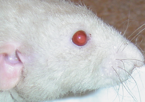 Photo 1: Right eye glaucoma. |
Case history and photo courtesy of Paige Anderson
Veterinarian: Dr. Bogan, DVM, Banfield Veteranary Hospital in Waterford Lakes Orlando

