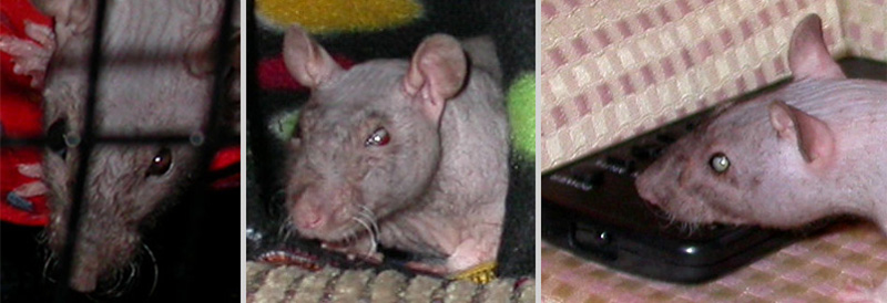
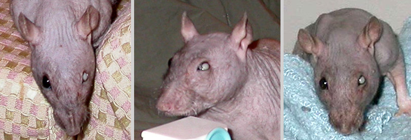
Figure 1: Anterior lens luxation in female rat (Gemma).
Case history and photos
History
Gemma was a rescued, double rex female rat with peach colored fuzz and ruby eyes. She was born in late August 2003. There was no reported history of previous illness. Gemma was housed together with three female cagemates.
Clinical Signs
In November of 2003 the owner noticed a reddish-white crescent-shape inside the left eye. Gemma did not exhibit any signs of pain or discomfort. An appointment with the vet was made, and she was seen within that week during which time the lens had popped out of place and fallen forward.
Diagnosis
Anterior lens luxation of undetermined cause.
Treatment
The vet determined that surgical repair/lens removal was of too great a risk in a rat, and that the condition could be medically followed in the event any secondary complications presented.
Outcome
By December 10th of 2003 the lens of the left eye was almost completely out of place, and by December 25th the lens had settled in to its final position at which time the eye began to shrink. No further changes occurred to the eye, and she adapted well with the just the use of her right eye. The condition never slowed down her activity, and she continued to enjoy running for hours on a rodent wheel every day.
Follow-up
In May of 2004, two of Gemma’s cagemates passed due to age related disease. Gemma showed close emotional attachment to her remaining cagemate Melanie. Two additional girls were adopted to become cagemates. However, when Melanie died in August of 2004, Gemma grieved to the point of illness and succumbed.
Photos
 In the photo to the left a crescent-shape can be detected in the left eye as the lens begins to fall forward. The center photo shows continuing displacement of the lens, and the photo to the right shows the lens is completely displaced. |
 Row 2 photos depicts the anterior lens displacement from 12/31/2003 to 3/18/2004 as the position of the lens settled. |
Case history and photos courtesy of M. Angel King


