Figure 2: Mononuclear Cell Leukemia in a 23-month-old rat (Lulu).
Case history and photos
History
Lulu was a 23-month-old female rat with no history of illness.
Clinical Signs
For about one month this rat had an abdominal mass with no other signs of illness. End stage clinical signs included: shallow raspy breathing, extreme lethargy, glassy eyes, pale extremities and low body temperature. Palpitation revealed that the “abdominal mass” was severe splenomegaly.
Diagnosis
Lulu was not diagnosed while she was alive.
Treatment
N/A
Outcome
Euthanized.
Follow-up
NECROPSY
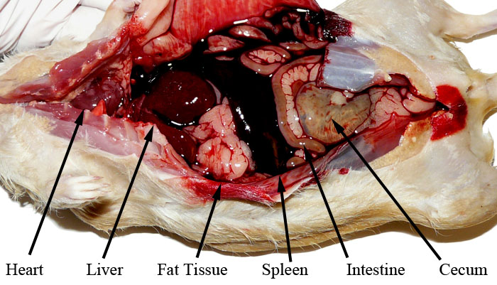
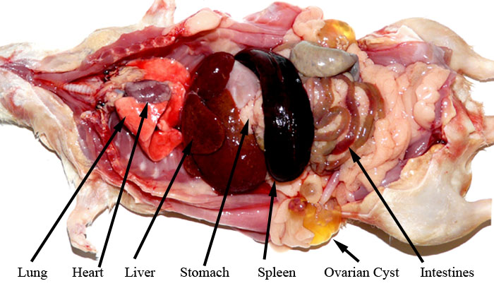
The abdominal cavity was filled with blood. The spleen was enlarged and ruptured. It is not known if the rupture occurred naturally or during palpitation. Incidental findings were bilateral ovarian cysts as well as a pituitary adenoma.
DISCUSSION
Extensive histopathology showed acute mononuclear cell leukemia with splenic rupture. Mononuclear cell leukemia originates outside bone marrow (lymphoid leukemia). This is the most commonly found leukemia in rats and typically originates in the spleen.
Photos
 In the above left photo you can see where the enlarged spleen has caused a noticeable widening of Lulu’s mid-section. The photo on the right shows the spleen removed with the rupture clearly visible. A normal rat spleen is about 1/10th the size of this one. |
 The above labeled photograph shows the internal organs in place and the abdominal hemorrhage. The spleen is transversing the abdomen. The spleen’s natural position is dorsal to the stomach which would hide it from view at this point in a normal necropsy. |
 The above labeled photograph shows the internal organs in place after the blood has been rinsed from the cavity showing the splenomegaly as well as the splenic rupture. The stomach, heart, and lungs appear normal. The liver is mottled and discolored from the leukemia. The intestines show advanced decay which may be attributed to natural post postmortem breakdown. The ovarian cysts are clearly visible. |


