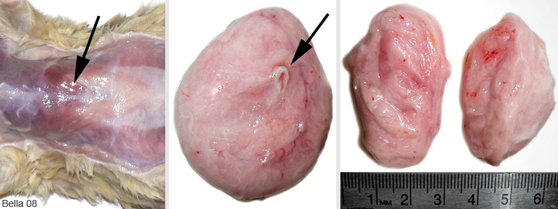
Figure 3: Lipoma in a 30-month-old male rat (Badger).
Case history and photos
History
Badger, a 30-month-old intact male rat with a history of respiratory issues.
Clinical Signs
For the last several months of his life the rat presented with a soft slow growing subcutaneous mass in the area of the ventral thoracic region.
Diagnosis
A fine needle aspirate was performed and the mass was identified as a lipoma.
Treatment
Due to the rat’s advanced age and poor health it was decided not to surgically remove the mass. Since the mass was benign, non-invasive, exhibited a slow rate of growth, and did not interfere with the rat’s daily activities it was not viewed as a critical surgery.
Outcome
Badger died from respiratory illness at 30 months old. The necropsy shows a typical benign lipoma with minimal attachment. Had the rat been a candidate for surgery this would have been a simple tumor removal.
Photos
 |
|
| Photo 1: Postmortem view of the subcutaneous mass. | Photo 2: The lipoma is exposed when the skin is peeled back. |
 |
||||
| Photo 3: After the lipoma has been removed you can see the minimal attachment to the underlying tissue (arrow). | Photo 4: The dorsal view of the mass shows the point of attachment (arrow). | Photo 5: The bisected lipoma shows scant vascularity. | ||
Case History, necropsy, and photos by J. “Bella” Hodges with the owner’s permission.


