Figure 1: Epitheliotropic lymphoma (also known as Mycosis fungoides) in 24-month-old rat (Prin).
Case history and photos
At one time this skin cancer was diagnosed as a fungal infection. Now it has it’s real name: epitheliotropic lymphoma. It is a rare type of skin cancer and very hard to treat.
History
Prin had a mammary tumor removed at the age of 15 months. She was also spayed. At 24 months Prin was in perfect health, never had myco related or other respiratory problems.
In the beginning of March of 2004, Prin developed what seemed to be a very small lesion. It was slightly swollen. A couple of days later, the swelling went down and the skin began to scab. The scab started to grow bigger, including more skin surface. A couple of weeks later, the scab was the size of a nickel. By the time we had the vet’s appointment on March 25th, Prin had another lesion growing on her side.
Diagnosis
On initial exam, suspected allergies.
Biopsies taken from 4 sites two weeks later shows Epitheliotropic Lymphoma.
Treatment
At the initial exam she was prescribed and placed on cefadroxil for two weeks and housed in an aquarium tank as opposed to a cage. The cefa drops helped in that the scabs did not spread rapidly, however many more scabs were formed. Two weeks later, the vet took 4 biopsies, and at this time the skin looked to be necrotizing (rotting). In the two weeks that it took for the results to come in, Prin’s body is almost completely covered in scabs. Her head and her back end is not affected at this point. Scabs began forming on her stomach. The biopsies taken two weeks prior revealed Epitheliotropic Lymphoma.
April 15th: Prin was started on steroids (Novo-Prednisone) to control the itching and pain. Dr. Venneval, the vet treating her, did some research regarding the use of Accutane. We will soon be starting the treatment. It has been said to reverse the affects of this cancer in some ferrets. Prin is showing signs of pain and discomfort. She is looking very old and slow. She still loves to eat and will still give you kisses.
April 23rd: on this visit to the vet, we have decided to wean Prin off of the steroids and added cefadroxil to her med list. She has started the Accutane (Isotretinoin). The reason for the cefadroxil is because the scabbing is starting to crack revealing her insides. This is causing Prin much pain. During the first week on Isotretinoin, she was taking 0.01 mL. There were no changes to the scabs or lesions on her skin.
April 29th, 2004: Prin was put on a higher dose of Isotretinoin: 0.02ml. But within a couple of days, it was decided this was too high (loss of weight). So it was reduced back to 0.01 mL. Her antibiotic was changed to enrofloxacin, and she was given Metacam as it was clear that Prin was in pain. We also started hydrotherapy. It flushed away a lot of the scabs, dead skin to reveal fresh new skin, but in a couple of hours, that skin began to scab over once again. Also, Prin developed scabs on her face, stomach, and back end. She was basically covered in either scabs or red crusting spots. The hydrotherapy caused her a lot of pain when her body started to dry. She was no longer sleeping, she couldn’t lay down in a comfortable spot. She was also having spasms of pain constantly.
Outcome
May 12th: we discovered Prin’s genital area was scabbing over causing her extreme pain. We decided in Prin’s best interest on euthanasia. She was assisted in passing quietly from us this day.
Follow-up
Regarding Prin’s necropsy result: Good news was her organs were not damaged by the Accutane. But bad news, Accutane at the dose we were giving her did not help whatsoever. In fact the cancer had begun attacking the muscles and surrounding tissues which was the cause of Prin’s spasms all the time.
Photos
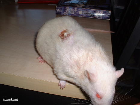 Photo 1: Single presenting lesion. |
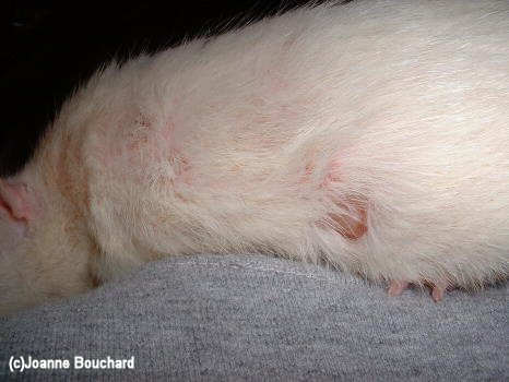 Photo 2: Single lesion with other presenting excoriated areas. |
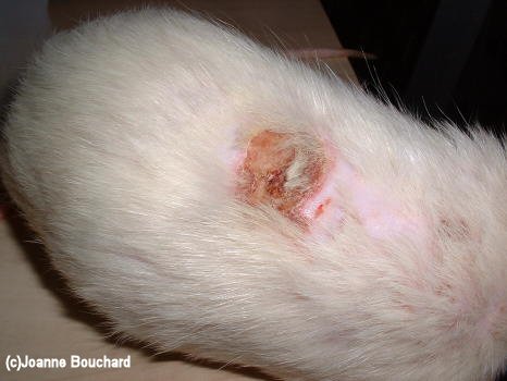 Photo 3: Lesion with notable area of alopecia (hair loss) |
 Photo 4: Scabbed and excoriated areas. |
 Photo 5: Scabbed lesion with more observable excoriation developing. |
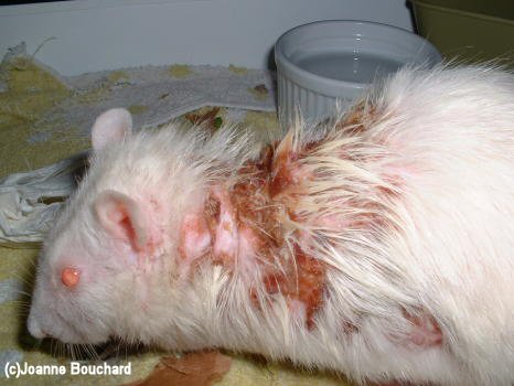 Photo 6: Scabbed, excoriated areas, and alopecia progressing. |
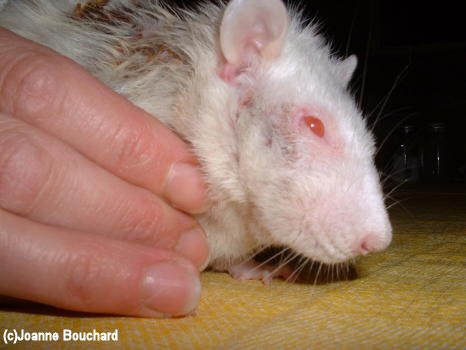 Photo 7: Facial involvement. |
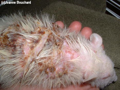 Photo 8: Disease progressing. |
 Photo 9: Disease progressing. |
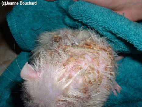 Photo 10: Disease progressing. |
Case history and photos courtesy of Joanne Bouchard


