Figure 4: Immunoblastic lymphoma with metastasis in 18-month-old male rat (Apollo).
Case history and photos
History
Apollo was an 18-month-old Siamese male from an immunodeficient line. Apollo weighed 548 g.. He was treated for chronic respiratory issues and had a melanoma removed from his right ear at approximately 10 months of age.
Clinical Signs
A mass formed on his neck and grew rapidly. Over a period of about a month the mass increased in size to the point where he had trouble swallowing and breathing.
Diagnosis
Cervical cancer- undetermined.
Treatment
Surgery was not an option due to his poor respiratory condition. Palliative care was provided. The rat was already on steroids and antibiotics for his respiratory condition. Soft food was given to make swallowing easier.
Outcome
When the mass became so large that he had to hold his head upward to breathe, the owner chose euthanasia.
A necropsy was performed, and tissue was sent to IDEXX RADIL for histopathology.
Follow-up
NECROPSY
Two masses were removed from the rat’s cervical area. Salivary glands were attached to both of them. Multiple white lesions were noted on the liver and the lungs. A mass was also noted within the spleen.
HISTOPATHOLOGY
SUMMARY
The cervical masses attached to the salivary glands are consistent with immunoblastic lymphoma. The lymphoma cells were also found multifocally in the spleen, lungs, and liver.
Cervical masses: Immunoblastic lymphoma characterized by aggregates of noncohesive 7-12 micron pleomorphic neoplastic mononuclear cells with round to oval, occasionally cleaved vesicular nuclei, moderate to abundant pale eosinophilic cytoplasm and 8-10 mitotic figures per 400X.
Liver: Immunoblastic lymphoma
Lung: Immunoblastic lymphoma
Salivary gland: No significant lesions
Spleen: Immunoblastic lymphoma
Photos
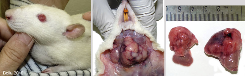 In the first photo, on the left, you can see how large the mass has become. The middle photo shows the mass exposed during necropsy. Beneath the mass is the esophagus and the trachea which is why breathing and swallowing became difficult. In the last photo the mass has been removed and you can see that it is actually 2 separate masses. Both are attached to normal salivary glands. The salivary glands are below each mass in the photo. |
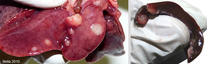 The photo on the left is of the liver showing multiple pale masses The photo on the right is of Apollo’s spleen which also has masses. Histopathology results showed that the masses on the liver, spleen, and the lungs (not shown) were a result of metastasis of the immunoblastic lymphoma. |
Necropsy and photos by: J. “Bella” Hodges
Histopathology by: IDEXX RADIL (University of Missouri)

