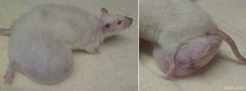
Figure 3: Large mammary tumor in female rat
Case history and photos
History
Female rescue rat 2.5-years-old.
Clinical Signs
Large mass located in the caudal region. Thinning fur, wasted appearance, along with insatiable hunger.
Diagnosis
Undetermined type of mammary tumor. Suspected to be fibroma.
Treatment
Veterinarian informed owner that surgery was not an option, due to location and size of mass (then about the size of a ping pong ball), and the age of the rat. Same vet advised the owner not to euthanize until the rat refused fluids and food.
Outcome
Humane euthanasia was sought and provided at a different veterinary clinic.
Follow-up Discussion
This case history provides a good example of what happens if an excisable tumor is not removed and allowed to grow indefinitely. Mammary tumors in female rats are common. The majority of which are benign fibromas that can usually be removed even in the are of the groin (caudal region), as long as the surgery is done before the tumor becomes too large and involved.
It is noted that by the time this rat was seen, by the veterinarian, he felt that surgery was not an option. Because of the assessment and recommendation by the owners initial veterinarian, the rat never did get euthanized by the owner’s vet since she continued to eat and drink. Almost all of the nutrients she consumed were being used by her body to sustain the fast growing mammary tumor. She ate ravenously right up till the day she was finally taken to another vet and euthanized. She was literally starving to death no matter how much food she consumed.
Although it may be the general rule of thumb, “as long as the rat is eating and drinking, wait to euthanize”, there are times where this is not going to apply. In a circumstance such as the case history described above, euthanasia would be the most humane course. Mobility for this rat was severely limited, the rat was constantly in a state of extreme hunger, and the tumor had begun to die and become necrotic in the area farthest from the blood supply.
During necropsy of this female, the tumor was removed and weighed. It was 9.2 ounces which is more than the weight of the 8.5 ounce rat. The body was totally devoid of any fat deposits, which are common in rats. The internal organs were small and exhibited signs of degeneration, atrophy, and disease. Her organs were literally rattling around in her body cavity. The rats stomach was filled with a large amount of food which showed her attempt to alleviate her hunger. The stomach also had a lot of air, indicative of either respiratory distress or pain (rats will swallow air when in pain).
Photos are graphic
 In the photos above you can see the rat is wasting away. Note the thin appearance of the neck, face, and back. The photos also show how far this tumor spreads from the outside of the rats right leg, below the rat, and under the left leg making walking impossible. |
 In the photo to the left you can see the underside of the tumor and the area that is necrotic. The second photo to the right, shows the tumor after it has been removed and spread out, and how it compares in size to the rat. |


