Figure 1: Mast cell tumor in 1-year-old male rat (Griffen).
Case history and photos
History
Griffen a 1-year-old intact male rat. Housed with male siblings.
Clinical Signs
We noticed a slow growing lump on his abdominal region in the lower left quadrant. To the touch it was hard, flat, and seemed to be unattached. When the lump reached about an inch in diameter it developed a bright red round raised “lesion” on top of it about 1/2 inch in diameter. The surface lesion was moist to the touch but not actively bleeding. I would describe it as similar to an ink stamping pad that would only ooze a small amount of fluid when pressed against something.
Diagnosis
On evaluation by the veterinarian it was determined that the lump was a tumor.
Due to the shape and the behavior of the growth the veterinarian speculated that it was most likely a “button tumor” (term use for histiocytoma or in some cases mast cell tumor).
Treatment
The rat was prescribed 1 mg orally of prednisolone once a day to help to shrink the tumor. After a week on the steroids there was no noticeable difference.
The rat’s overall health was good so the decision was made to surgically remove the growth. Mast cells are large connective tissue cells that contain granules filled with histamines, heparin, and seratonin. Due to the likelihood of histamine release during surgery the rat was also given a small amount of Children’s Benadryl at 0.2 cc twice a day for several days before the scheduled surgery date.
It was speculated that the red lesion on top of the tumor was either a local reaction to the compression of the normal surrounding tissue or a result of histamines being released resulting in a single welt or large hive.
Outcome
The surgery was successful. The vet removed as much surrounding tissue as possible to reduce the chance of any cancerous cell remaining. Due to the length of the incision “mattress” stitches were used in addition to the surgical glue for closing up the site.
Followup
The rat was kept on antihistamines for several days post-op.
The tumor was put into formalin and sent to the lab for histology.
The result of the laboratory histology report is as follows:
-
“The mitotic index is low meaning this is not an aggressive growth, and likely not malignant. The tissue is: not infiltrated, homogenous, it is encapsulated, well circumscribed (not invasive), and likely benign. The special stains for mast cell tumors (toluidine blue and giemsa) were not very useful because there were not many granules. Rodents may not have very granular mast cell tumors. I am unable to tell you definitely if it is mast cell tumor, but there were granules present, so it is possible. But highly likely not malignant and self contained. The vet should have been able to remove the entire tumor since it was well contained and not invasive to the surrounding tissues. ”
The prognosis for this rat is very good. As a safeguard this rat will be checked often for signs of any more growths.
Photos
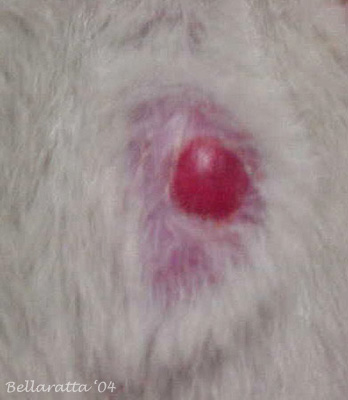 Photo 1: Close up view of mast cell tumor |
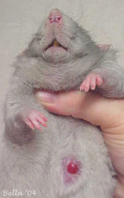 Photo 2: View of mast cell tumor located on the abdomen |
Case history by Joanne “Bella” Hodges
Histology report by Charlene M. Crain, MBA, BSMT (ASCP)

