Figure 3d: Obstructive Megacolon due to Fibrosarcoma
Case history and photos
History
This is a 16 month-old female rat, Tinkerbelle.
Clinical Signs
She presented with a severely distended, hard, abdomen and weight loss. Her spine and hip bones protruded, indicating that she was malnourished. The owner had seen no evidence of the passage of stool for a week. She did state that the rat still had a good appetite.
Diagnosis
Obstructive Megacolon
Treatment
None.
Outcome
Humane euthanasia.
Follow-up
A necropsy was performed and tissue was sent to IDEXX RADIL for histopathology.
NECROPSY
The necropsy revealed: severe megacolon, thickened rectal tissue, and rectoanal lesions obstructing the passage of stool.
HISTOPATHOLOGY
SUMMARY
A portion of the terminal colon and rectum with anus from a female, 16-month-old, rat was submitted due to an enlargement of the intestine just proximal to the anus. The mass is consistent with a fibrosarcoma.
Anal Mass: An unencapsulated mass of spindle shaped cells with large vesicular oval to round nuclei, with some nuclei being smaller and condensed, with pale blue to moderately eosinophilic strap-like cytoplasm. An eosinophilic fibrillar
refractile material is being produced by some of the cells. This material is consistent with mature collagen. The cells are woven in interlacing bundles in some areas and other areas are more solid with less of this eosinophilic material being produced. There are large areas of cellular necrosis and occasional areas of the mass where the cells are very loosely arranged and separated by a mucinous ground substance. Mitotic figures are not very common even at 400X magnification. The anal epithelium is regionally ulcerated with numerous bacterial colonies present on the exposed dermis. The mass is consistent with a fibrosarcoma.
Necropsy Photos
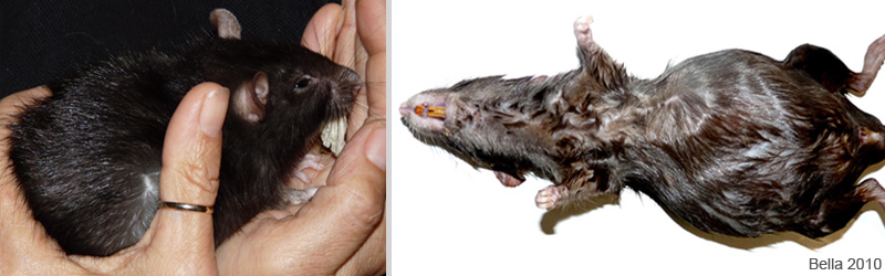 The photo on the left shows Tinkerbelle before euthanasia with a biscuit in her mouth that she would not let go of. One of the issues with megacolon is that the colon cannot properly process food. Even though Tinkerbelle had been eating, she was actually starving resulting in failure to thrive. In the photo on the right she has been wet down prior to the necropsy, and you can see the severity of the abdominal distention. |
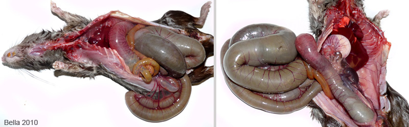 The photo on the left was taken immediately after the abdomen was cut open. The intestinal tract was under so much pressure that it literally popped out of the body cavity. The photo on the right shows a greatly enlarged colon lifted away from the body. Note the complete lack of internal body fat. |
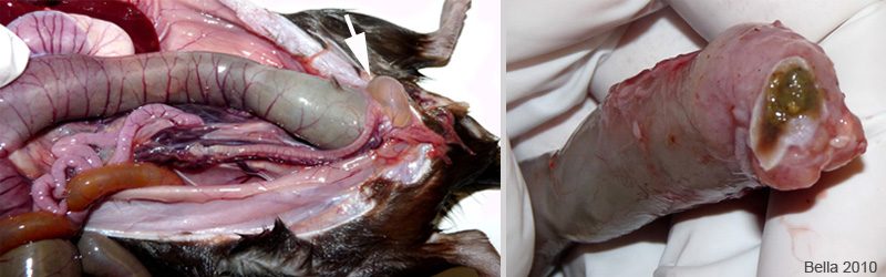 The photo on the left shows a close up of the descending colon and the rectum. The white arrow points to the urinary bladder. The blocked rectum is to the left of the bladder. The photo on the right shows a close-up of the rectum and anus after being removed. The wall of the rectum, proximal to the anus, is thickened and discolored. |
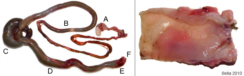 The photo on the left shows the lower gastric structure removed, cut free of the mesentery membranes, and labeled. A. Stomach, B. Small intestines, C. Cecum, D. Colon, E. Rectum, F. Anus. The photo on the right is a close-up of the rectoanal section after it has been cut open and rinsed. One can see how thick and nodular the affected tissue had become. |
Case history, photos, necropsy: J. “Bella” Hodges
Histopathology by IDEXX RADIL

