Lipoma, cystic thoracic lymph node, and nephropathy in an aging male.
Case History
A 28-month-old male fawn dumbo rex rat. Clinical signs of illness at time of euthanasia: severe hind end paresis, mild breathing difficulties. Weight: 532 grams (1 lb. 2.8 oz.)
Necropsy Pictorial
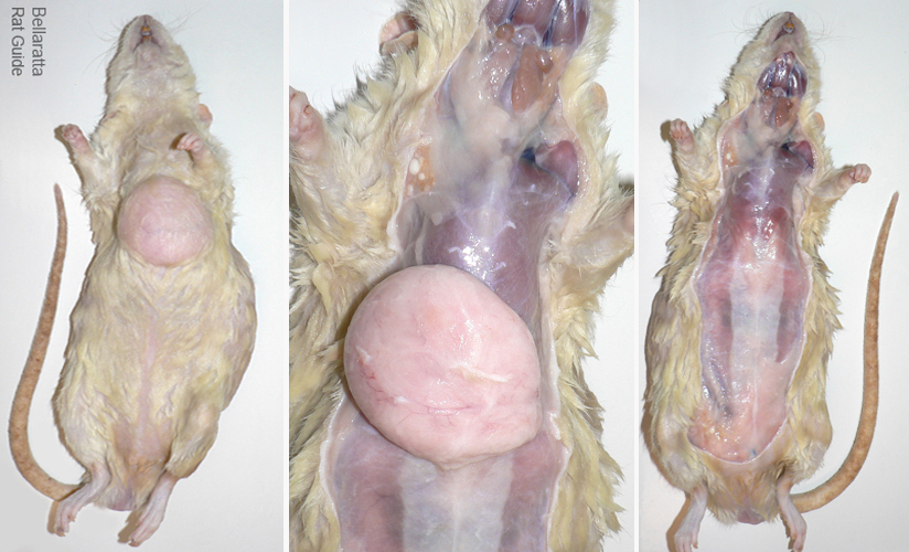
 From left to right, the first photo shows the rat prepared for the necropsy. The dampened fur makes the mass easy to see. In the middle photo the skin is pulled back and the lipoma is exposed. The photo on the right shows the torso after the tumor has been removed for examination. |
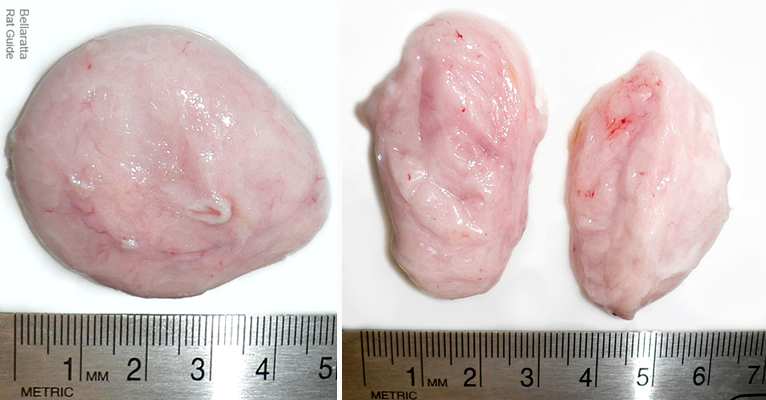
 Note the lack of a major blood supply to the tumor. Lipomas are slow growing benign tumors composed of fatty (adipose) tissue encased in a thin membrane of connective tissue. Subcutaneous lipomas are located between the skin and the muscle. |
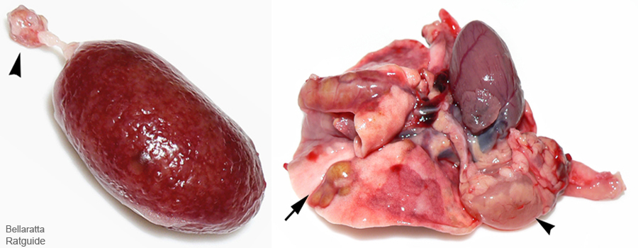
 The photo on the left is a kidney. The arrow head is pointing to the adrenal gland. There is marked pitting on the kidney surface: This is often an indication of chronic nephropathy (kidney disease). Some of the factors that can contribute to kidney disease are: aging, long term use of certain medications, a high calorie diet (especially combined with high protein), hypertension, and diabetes. The kidneys are the first organs to decline in age. Male rats, due to a unique urinary globulin, are particularly susceptible to renal degeneration. The right photo is the thoracic pluck. The arrow shows a pulmonary abscess typical of bacterial infections such as Mycoplasma pulmonis. The arrowhead shows a cystic thoracic lymph node |
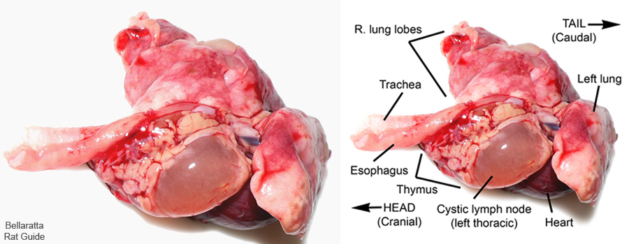
 The photos above are of the thoracic pluck. The fluid filled sac is a dilated lymph node most likely due to obstruction of the lymphatic ducts somewhere “upstream” in the lung. The parts have been labeled for easier identification. |
Necropsy and photos by Joanne “Bella” Hodges
Consultation: Research Animal Diagnostic Labortory (IDEXX RADIL)


