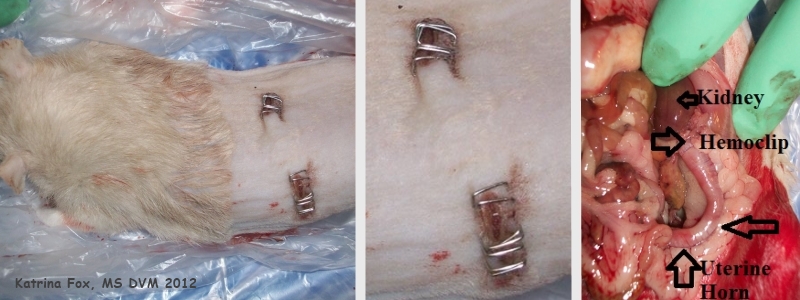
Figure 1a: Ovariectomy in 1-year-old female rat (Octopus).
Case history and photos
History
“Octopus” was an approximately one year old rescue rat of unknown background. The decision was made to do an ovariectomy, in order to reduce the chance of her developing additional mammary tumors or a pituitary tumor.
Clinical Signs
At the time (February 2012), Octopus had one mammary tumor.
Diagnosis
Apart from the mammary tumor, Octopus appeared to be in good health during her pre-op exam.
Treatment
Octopus was given Buprenorphine (painkiller) pre-op. Isoflurane was used for anesthesia.
The mammary tumor was removed. In addition, an ovariectomy (OE) was performed. The ovaries were removed through the back wall of the abdomen. The two incisions in Octopus’s back were closed with Autoclips (brand name, “staples”).
Octopus was given Metacam post-op and placed in an incubator with oxygen.
Outcome
Octopus appeared to be doing fine, then went into respiratory distress and passed away during the recovery.
A subsequent necropsy indicated that there had probably been “some pre-existing respiratory disease that was subclinical at the time”.
Photos
 The two (postmortem) photographs on the left show the use of Autoclips (brand name) to close the two incisions that were made in Octopus’s back in order to perform the OE spay. The photograph on the right was taken during the necropsy; showing the lower abdomen. The arrows indicate the uterus (which remains in situ during an OE spay), the left kidney, and a Hemoclip. |
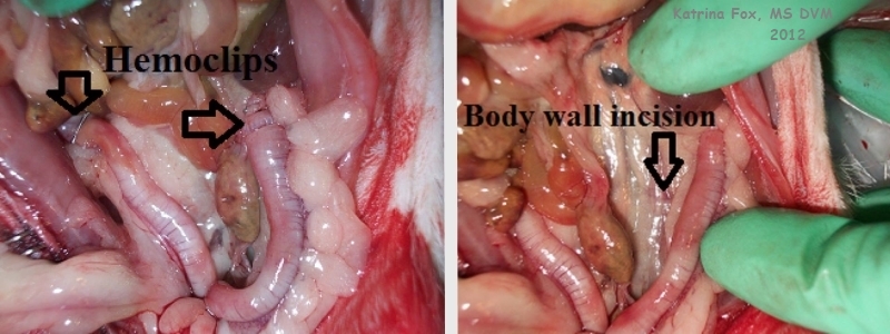
 These necropsy photographs also show the lower abdomen. The photograph on the right provides an internal view of the incisions that were made in Octopus’s back for the OE spay. |
Case history and photos courtesy of: Katrina Fox, MS DVM
Case Editors: Karen Grant RN, and Cyzahhe


