Figure 2: Pyometra in a 24-month-old hairless female rat (Cricket).
Case history and photos
History
Cricket is a 24-month-old hairless female rat.
Clinical Signs
2/5/05
Cricket began showing signs of bleeding from her vaginal area. She was started on antibiotics to get her through the weekend.
2/7/05
Cricket had her first veterinary visit. She continued to show signs of bleeding in the vaginal area.
Diagnosis
This vet reported infection due to genital mycoplasma
Treatment
Cricket was started on Baytril. Within one week a small growth appeared in her groin and a large one on her back (see photos below).
The one on her back doubled in size quickly, and a needle biopsy showed no fluids. The veterinarian kept with his original diagnosis, and we sought a second opinion.
Follow-up
2/18/05
Cricket was seen by another veterinarian for a second opinion. This vet began Cricket on a Baytril/Panmycin cocktail to be sure to rule out infection. Cricket responded well to this treatment, and the bleeding stopped temporarily.
3/3/05
Cricket turned two years of age and began to start bleeding again. We took her to the vet again on 3/4/05. The vet believed the best option at that point was surgery. He agreed to remove the mass in her groin and to do some explorative surgery to determine the cause of her bleeding.
3/7/05
Cricket underwent surgery. The vet removed the mass from her groin and found her uterus to be diseased. He spayed her. She was under anesthesia for over one hour. The incisions required 14 sutures to her stomach, and 2 to her groin. She was remarkably smaller in size following removal of the growth and uterus. The vet compared the enlarged uterus to the size of a cat’s. He also noted that the mass appeared to be filled with blood. Biopsy of both the mass and her uterus were sent to pathology.
Outcome
Cricket’s recovery at first was slow. She refused to drink fluids initially, and it took several days of persistent nursing care for her to become her active self again. We are hoping with a full and extended recovery that she may be able to go back and have the growth from her back removed.
Following the removal of Cricket’s sutures, she showed signs of pus draining from her vagina and was started back on Baytril.
Cricket fully recovered from her surgery and follow-up treatment.
Pathology Report: Cricket
Gross: Received 2 slides labeled uterus and lumbar mass, 4 x 1.2 and 1 x 1.
LUMBAR MASS MICROSCOPIC: The specimen consists of presumptive mammary tissue with a mass composed of proliferation’s of myoepithelium and glands lined by a single layer of plumb but monomorphic epithelium. The lesion extends to margins on the section examined.
DIAGNOSIS: Fibroepithelial hyperplasia.
COMMENT: This is a benign lesion that may be the result of hormonal stimulation. Excision of the mass, and removal of hormonal stimulus should be curative, but follow-up is advised as malignant tumors may arise from sites of hyperplasia.
UTERUS MICROSCOPIC: The specimen consists of uterus with variably thickened endometrium with infiltrates of macrophages and plasma cells and cystic glands containing neutrophils and macrophages.
DIAGNOSIS: Cystic endometrial hyperplasia with endometritis.
COMMENT: This is compatible with pyometra. Culture is recommended to look for infectious agents.
AR KIEHL DVM, MS, DACVP
Photos
The following are pre and post-op photos of Cricket.
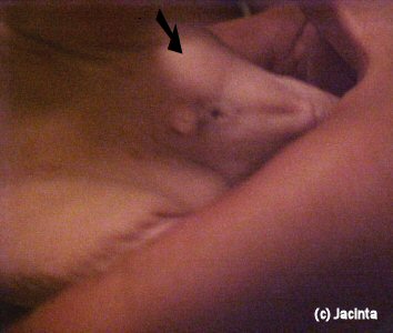 Photo 1: Mass in groin indicated by arrow. |
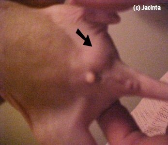 Photo 2: Additional photo of mass in groin indicated by arrow. |
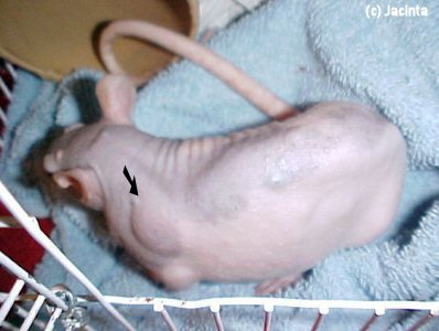 Photo 3: Post-op day 1. Also showing mass on back indicated by arrow. |
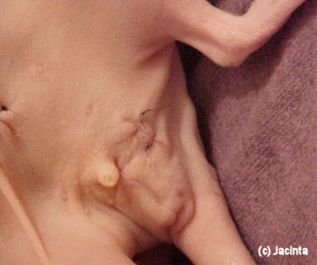 Photo 4: Sutures at site of groin mass. |
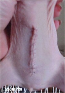 Photo 5: Post-op day 1 showing suture site after removal of pus-filled uterus. |
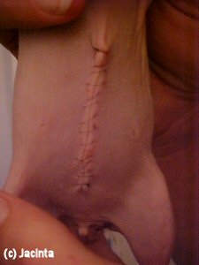 Photo 6: Post-op day 2 showing suture site after removal of pus-filled uterus. |
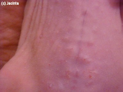 Photo 7: Shows healing complete. |
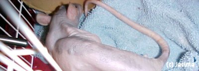 Photo 8: Shows decreased weight and size following removal of uterus. |
Case history and photos courtesy of Jacinta Sousa.

