Figure 3: Pyometra and subsequent intussusception in a 12-month-old female rat (Lily).
Case history and photos
History
Lily is a 1 year old intact female rat.
Clinical Signs
Developed vaginal bleeding.
Diagnosis
The vaginal bleeding was suspected to be due to pyometra.
Treatment
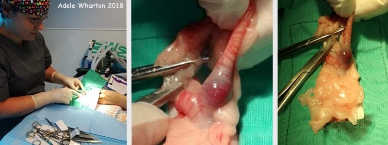
Lily was spayed. During the surgery it became clear that one of the uterine horns was indeed affected by pyometra: it was filled with pus and blood. In addition, the cervix was noted to be abnormally edematous, which was also thought to be due to the infection.
While recovering from the surgery, Lily went into arrest. She was successfully resuscitated by administering chest compressions, oxygen and adrenaline.
Lily received meloxicam pre-op and buprenorphine on recovery. In addition, Lily’s veterinarian always administers local anesthesia (lignocaine splash block) after muscle closure in spay or other abdominal surgeries. Meloxicam was continued post-op for 5 days.
Follow-up
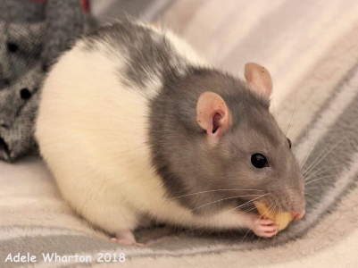
Lily initially recovered steadily at home over the next couple of weeks, and was successfully reintroduced to her cagemates.
Within 2-3 weeks following the surgery, however, Lily once again started showing (intermittent) vaginal bleeding. Over the following weeks she received several courses of antibiotics (enrofloxoral, clavamox, clindamycin), however, she was unresponsive to the antibiotics and the vaginal discharge continued, varying from frank (bright red) bleeding to clotted blood to purulent blood. In addition, Lily’s caudal abdomen began to distend. During re-examination the veterinarian palpated a mass, within which the cervical ligature from the spay surgery could be felt. Ultrasound indeed showed a soft tissue mass, with fluid pockets in the region of the cervix, thought to be consistent with an abscess or hematoma.
Given that Lily arrested during recovery from her previous surgery, both the veterinarian and Lily’s owner were reluctant to put her through another surgery. A swab was taken of the vaginal discharge and cultured, however it only showed an E. coli that was sensitive to all the antibiotics that had been given during the previous weeks and it was concluded to be a contaminant. Hoping that the abscess/hematoma would eventually drain on its own, the decision was made not to intervene and simply observe Lily closely for any changes. Over the following two weeks the intermittent vaginal discharge continued. The abdominal distension, however, visibly reduced and a follow-up ultrasound confirmed that the distention was definitely smaller.
Over the course of the weekend following the ultrasound, Lily suddenly showed acute pain and her vaginal discharge increased. After consulting the veterinarian, Lily was given buprenorphine, however this did not decrease the symptoms of pain. When she was taken to the veterinarian on Tuesday evening she was lethargic, fluffed up, pale, and sucking in her sides. At this point the only remaining options were euthanasia, or surgery with a slim chance of recovery. Lily’s owner opted for surgery.
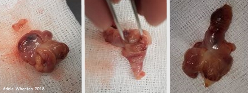
Lily was anesthetized with sevo/O2 and a caudal abdominal incision was made. The mass was immediately visible in the caudal abdomen, wrapped in omentum. As the invention omentum was dissected away, it became clear that the mass was adhered to the bladder caudally, the colon cranially, and both ureters on either side. Given the difficulty and risk associated with removing it without damaging the surrounding structures, Lily’s owner was consulted. The decision was made to continue with the very challenging surgery. Note that, at this time, the mass was thought to be a tumor. During dissection it became clear that the mass was associated with Lily’s cervix and was in fact a cervical intussusception, and indeed included the cervical ligature from the previous spay surgery, as had been felt by the veterinarian during the earlier examination. After separating the “mass” from the other surrounding tissues, it was separated from the vagina at the pelvic rim and successfully removed. The abdomen was then thoroughly flushed with saline and closed with a routine midline closure, and Lily was given local anesthesia.
Outcome
Unfortunately, during the recovery period from the surgery Lily arrested. She was successfully resuscitated and stabilized sufficiently to be transferred to an incubator/oxygen tent, however, she again arrested and this time the veterinarian was unable to revive her.
Pathology
The mass, on investigation, was a cervical intussusception, complete with the original cervical ligature from the initial surgery for pyometra.
Photos
 Photo 1: The photo on the left shows veterinarian Adele Wharton performing the initial spay surgery on Lily; the vet tech has her hand under the drape to monitor Lily. The second and third photo show the veterinarian pointing to the diseased tissue that was removed during the spay surgery; the accumulation of pus and blood in one of the uterine horns due to pyometra is clearly visible. |
 Photo 2: This photograph of Lily was taken during the initial successful recovery after the spay surgery, and before the second, exploratory surgery during which the cervical intussusception was discovered. |
 Photo 3: These photographs show the “mass” that was removed during the second, exploratory surgery. Investigation showed that it was a cervical intussusception, complete with the original cervical ligature from the initial spay surgery. |
Case history and photos courtesy of Faye Hollands-Meadows (owner) and Adele Wharton, BVSc, MRCVS, CertGP (F&L)


