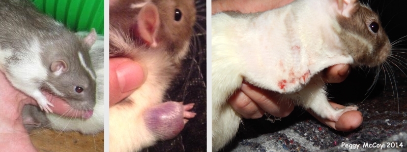
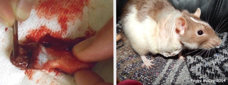
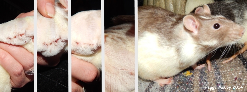
Figure 9: Soft Tissue Sarcoma of the forelimb and subsequent amputation in 22-month-old male rat (Ace).
Case history and photos
History
Ace (intact male) was born in April 2012 and adopted at 8 weeks of age. He lived in a large group of 20+ rats, which was fed Oxbow or Native Earth, with a daily portion of vegetables. Sugary treats were limited. Ace did not have any health problems until he was 22 months old.
Clinical Signs
Ace’s right hand appeared swollen. Metacam was administered for three days, at which time the swelling turned into what was thought to be a hematoma. The swelling was treated unsuccessfully with ice packs, warm compresses, and steroids. In the second week it was drained at the veterinary clinic, which revealed dark (old) blood. However, in the third week the swelling increased and a surgery was scheduled.
Diagnosis
Soft tissue sarcoma.
Treatment
On 28 February 2014 Ace’s right forelimb was amputated to just below the scapula.
He was given, and sent home with Metacam for pain relief as needed following recovery.
Outcome
When Ace was picked up from the veterinary clinic he was alert and walking around. He did not require further doses of Metacam. By the 6th day post-op his incision was almost completely healed.
Ace instantly adjusted to having only one forelimb. He quickly learned to eat with one hand and had no difficulty grooming or walking up the ramps in his cage. He was still able to keep up with the other rats and pin them if needed.
Follow up
Sadly, Ace passed away on 23 May 2014, only three months after his surgery.
Comment
Soft tissue sarcoma unconfirmed. No histopathology done on excised mass, but gross appearance appeared consistent for sarcoma.
Photos
 The photo on the left shows Ace’s right forelimb and hand before the swelling occurred. The photo in the middle shows the swelling. The photo on the right was taken immediately after the amputation surgery of Ace’s right forelimb. |
 The photo on the left shows the amputated right forelimb(orientation: the hand is on the left, underneath the tumor, which was cut transversely). The photo on the right was taken on day 1 post-op |
 These photos show the rapid healing of the incision. They were taken on, respectively, days 2, 3, 10 and 11 post-op. The final photo shows the completely healed incision site after regrowth of the fur. |
Case history and photos courtesy of Peggy McCoy


