Figure 2: Hemangiosarcoma in adult male rat
Case history and photos
History
Male, 14-month-old, agouti dumbo rat. No history of illness.
Clinical Signs
Owner noticed swelling on the back/side of the neck and extreme lethargy early in the morning. As the day progressed the swelling increased rapidly and he showed loss of motor control. The cervical mass felt hard and attached. The rat was taken to the vet in the late afternoon. His temperature was 95 degrees. He refused all food and fluids.
Diagnosis
A fine needle aspiration was performed at the veterinarian’s office and upon reading the slide cancerous type cells were seen. The rat was initially diagnosed with an unknown cancerous cervical mass.
Treatment
The rat was given oxygen and put on a heating pad to raise body temperature. He received an injection of Baytril. Rat died at the veterinarian’s office.
Necropsy and Final Diagnosis
A necropsy was performed on the rat and showed a large maroon cervical mass as well as maroon spotting on the lungs. The tumor, once removed and dissected, revealed a lumpy, dark red, gelatin like interior. The tumor and the lung tissue were put in formalin and sent out for pathology.
The findings showed that both the mass and the spotting on the lungs were hemangiosarcoma, an aggressive malignant cancer that originates in the cells that line the blood vessels (endothelial cells).
Discussion
Although it can theoretically occur anywhere blood vessels are present in the body hemangiosarcoma is most prevalent in the heart, on the skin (or under the skin), and in the spleen. Sarcoma, cancer of the soft tissue, is typically a highly malignant type of cancer. Even the skin forms tend to metastasize to other parts of the body.
Photos
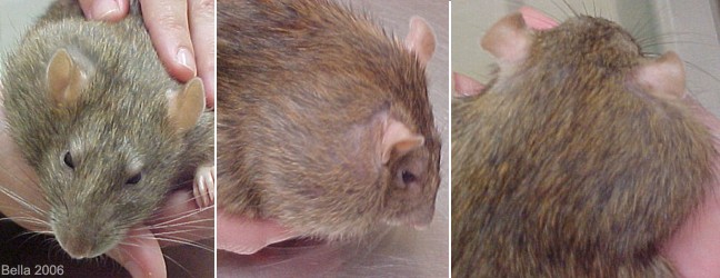 In the first photo on the left the rat shows a mass in the right cervical/shoulder region. The next two photos show the mass greatly enlarged only 6 hours later. |
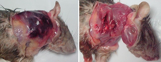 During the necropsy removal of the skin revealed a large dark red mass. Once removed the attachment was noticed to be at both the shoulder as well as the neck. |
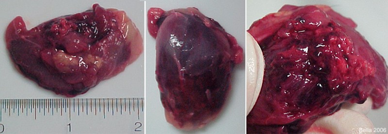 The first photo on the left shows the large size of the hemangiosarcoma (bottom of tumor shown) The middle photo shows the top of the tumor after removal. The photo on the right shows the inside of the tumor after it was cut open. |
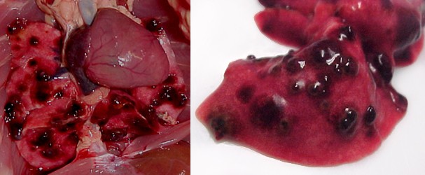 To the left is a photo of the lungs before removal from the body. The following photo shows a close up of a lung lobe with multiple, dark, and raised hemangiosarcoma tumors. |

