Figure 5: Squamous cell carcinoma of Zymbal’s Gland in female rat (Lauren).
Case history and photos
History
Lauren, a 20-month-old female rat with no history of illness.
Clinical Signs
Swelling was noted on the left side of the rat’s face, between and below her ear and eye.
Diagnosis
A fine needle aspiration revealed pus. Her condition was diagnosed as a large facial abscess.
Treatment
Lauren was masked down and the area was incised and flushed. A drain tube was inserted. The veterinarian prescribed high end dosage of Baytril. The facial area appeared much less swollen. The wound was flushed daily with diluted Betadine.
Outcome
The facial swelling worsened over the next week. She was taken back to the veterinarian to be reevaluated and to have the drain tube removed. At this point the swelling was so extreme that the skin could not be kept together with sutures.
Follow-up Diagnosis and Treatment
The veterinarian confirmed a diagnosis of facial tumor.
Lauren was kept on the Baytril and was administered an injection of dexamethasone. The veterinarian felt that the tumor was too invasive and aggressive for removal. Mild OTC pain relievers were administered for comfort.
Outcome and Pathology
Over the following week the mass grew rapidly. The pressure caused the jaw to shift and resulted in malocclusion. She still had a good appetite and continued to be active. Two weeks after the lump was first noticed Lauren started bleeding from her left ear and shaking her head. At this point the decision was made to euthanize her.
A necropsy was performed and tissue was sent to the lab. The final diagnosis: Squamous cell carcinoma of the Zymbal’s gland.
Photos of the Zymbal’s Gland Tumor
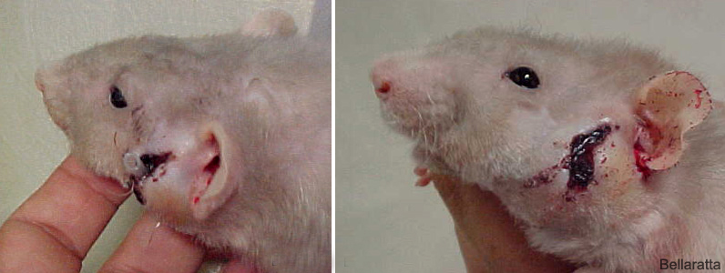 Above left photo shows Lauren with the drain tube in place. Several days after the “abscess” had been drained the mass quickly became apparent. In the photo on the right you can see that the enlarged mass prevented closure of the wound. |
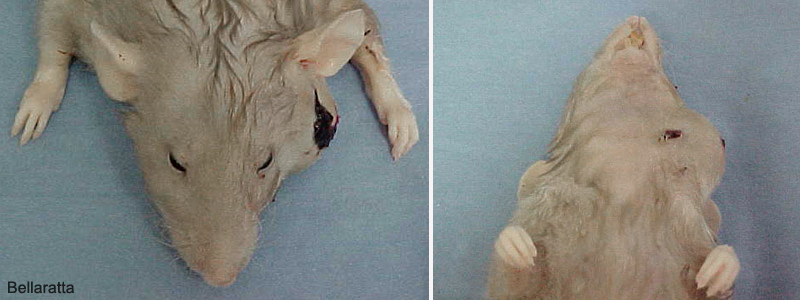 Dorsal and ventral post mortem views of the mass. |
 Necropsy photos showing the exposed tumor. |
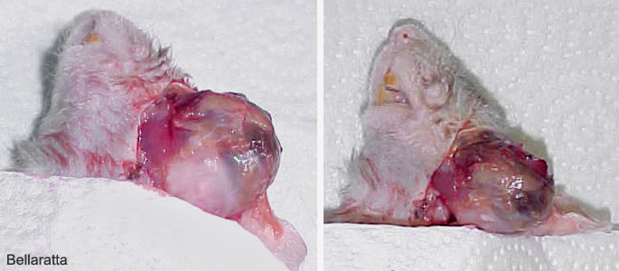
 The Zymbal’s gland tumor removed. In the left photo the area where the tumor was attached is shown. Due to the invasiveness of the mass only a portion of it could be excised. The photo on the right shows the distal portion of the tumor. These tumors grow very quickly. Only two weeks lapsed between the first sign of facial swelling to euthanasia |

