Figure 8: Fibroblastic tumors and incidental postmortem finding of Left Adrenal Gland Adenoma in a 31-month-old male rat (name: #7).
Case history and photos
History
A 31-month-old 597 g, SPF MYCO free, retired male lab rat, #7 (number name retained by owner), used in non-invasive behavioral experiments was adopted at 13 months of age. Abdominal neutering was performed shortly after being adopted. No other previous health history available.
Clinical Signs
Two small skin masses were noted. One mass presented on the right lateral thoracic area, and one on the left lateral thoracic area. Both were being closely monitored for growth. No increase or change was noted in the size of the mass on the left, but the mass on the right was increasing and the decision was made to have it removed.
Diagnosis
Left Dermal mass
Right subdermal mass attached to rib.
Treatment
Surgery was performed to remove the left dermal skin mass, and as much of the right subdermal mass with attachments to muscle and bone as possible. The surgery, and immediate recovery, was without incident. A body wrap was applied, and a post op injection of butorphanol was given to control pain.
Outcome
Following a successful recovery at the veterinary hospital he was transported on a two hour drive home. Upon arriving home he was alert and responsive, and readily ate some lentils and homemade chicken soup, along with drinking Ensure orally by syringe. Approximately an hour later his condition changed abruptly, identified (a) primarily by his eyes which bulged slightly and suddenly, giving the appearance of an increase in pain, and (b) in the way he stopped moving and lay there quite passively. As pain was suspected a dose of Metacam was given. The body wrap was removed immediately, and efforts to stimulate him were made on veterinary advice, including massaging him and pinching his hands and feet. He was non-responsive to all efforts. He also did not respond to additional offers of food and Metacam. Death ensued within 4 hours.
Follow-up
A necropsy with histopathology was obtained:
Biopsy Report of Left and Right mass:
DESCRIPTION:
- Left mass: This is a circumscribed mass partially covered by skin from the left lateral thoracic area. The mass consists mostly of an array of well differentiated fibrocytic cells with abundant eosinophilic collagen stroma. Several more cellular areas have atypical fibroplastic tumor cells with uneven sized nuclei and a few mitotic figures.
- Right mass: This is a non-encapsulated mass composed of long spindle cells with relatively even-sized oval vesicular nuclei forming streams and interlacing patterns. There is a dense stroma of collagen between the spindle cells. The mitotic activity is low.
FINDINGS:
- Left mass: Cutaneous fibroma with areas of atypia.
Comment: This is mostly a benign tumor of dermal fibrocytes with abundant collagen stroma and a few areas of atypia (pre-cancerous changes).
- Right mass: Cutaneous fibrosarcoma
Comment: This is a well differentiated spindle cell tumor with features of low grade malignancy. The tumor has some characteristics of fibrocytic origin (fibrosaroma).
These spindle tumors are often locally invasive but seldom metastasize. The surgical margins in the section submitted appear to be free of tumor cells; however there are some narrow deep margins.
Biopsy Report of Adrenal gland mass:
DESCRIPTION:
This specimen represents an adrenal gland which has been expanded and distorted by sheets and nodules of well-differentiated rounded cortical-type epithelial cells which have displaced normal structures of the cortex and medulla. These cells are generally well-differentiated and mitotic activity is not seen. The lesion has a background of hemorrhage and hemosiderosis with fibrin deposition.
FINDINGS:
Adrenal cortical adenoma with associated chronic hemorrhage and hemosiderosis.
Comment: Morphology is characteristic of a benign cortical adenoma which has deteriorated and caused hemorrhage. The lesion may have been related to this animal’s death if hemorrhage was extensive. Endocrinopathies may also have been involved.
Summary
Death unrelated to right and left fibromatous masses. The left adrenal gland was significantly enlarged– approximately 25 times normal size (see comparison to normal size right adrenal gland in photo below), and the artery to the gland was also greatly enlarged with a large clot formation.
Many of these tumors are functional and cause an overproduction of the chemicals normally made by the adrenal gland, bypassing the body’s normal ability to regulate the production. The report at necropsy indicates chronic bleeding occurred from the presence of the tumor in the gland.
Photos
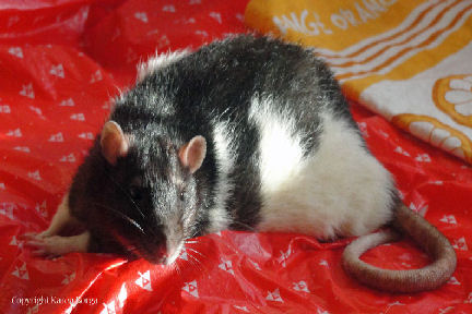 Left photo shows #7 prior to detection of masses. |
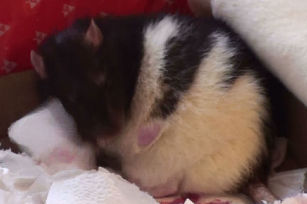 Right photo shows presence of left lateral dermal skin mass. |
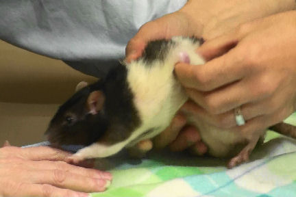 Left photo shows Dr. Pilny palpating for extent and depth of left lateral mass. |
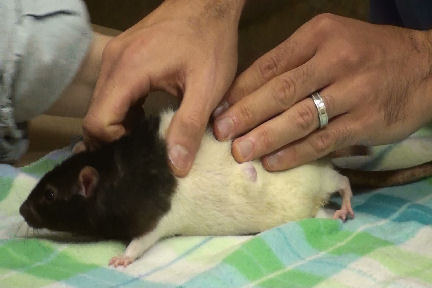 Right photo, again, shows palpation of left lateral mass. |
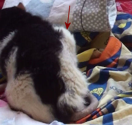 Left photo shows right lateral subdermal mass (indicated by red arrow). |
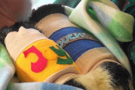 Right photo shows #7 postop following application of body wrap with blue Vetrap dressing. |
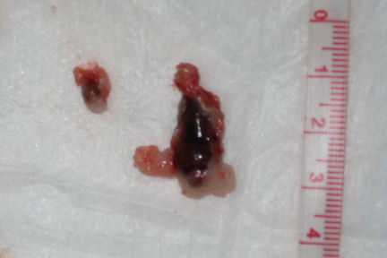 Photo shows the left (as seen on the right of the photo) enlarged adenomatous adrenal gland compared with the normal-sized right (as seen on the left of the photo) adrenal gland. The enlarged adrenal gland is 25x that of the normal adrenal gland. |
Case history and photos courtesy of: Anthony Pilny DVM DABVP, and Karen Borga
Necropsy and Summary: Anthony Pilny, DVM,DABVP
Histopathology report of left and right mass: R. Montali, DVM, Dipl, A.C.V.P, Dip., A.C.Z.M
Histopathology report of adrenal gland mass: C. McLeod, DVM, Diplomate A.C.V.P.

