Removal of large and small mammary tumors from a 2.5 year old female rat.
Complete procedure duration: 60 minutes
Surgery performed by:
Dr. Peter Field B.V.S.C. (Hons.) M.A.C.V.Sc.
Member of the Australian College of Veterinary Scientists by examination in Canine Medicine
Veterinary Physician and Surgeon, Everton Hills Veterinary Surgery, Everton Hills, BrisbaneQld, Australia
and
Surgical Assistant:
Mrs Louise Dux, Certified Laboratory Animal Technician
*Note: While it is Dr. Peter Field’s preference to use an approved surgical hand scrub, rather than surgical gloves to maintain aseptic technique with small animals, his technique does not replace the accepted, evidence-based, surgical practice of the need for the use of sterile gloves, or appropriate draping when performing surgery on rats.
Click on a photo to enlarge it.
tumour_removal_01
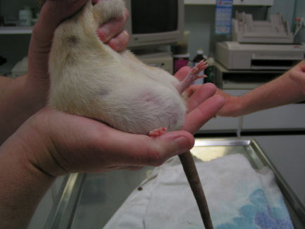
A 2.5 year old intact female rat brought into clinic for removal of small mass in the left thoracic area and large mass in the right lateral abdominal region.
tumour_removal_02
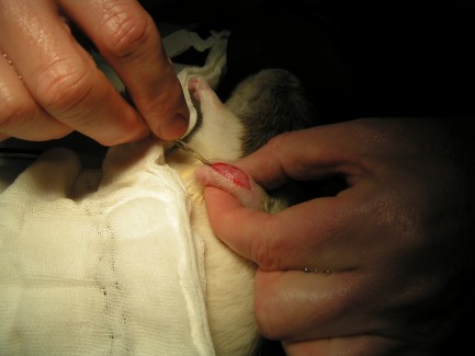
Smaller mass seen in the left thoracic area below forelimb.
tumour-removal-03
Larger mass seen at right lateral abdominal region.
tumour_removal_04
The rat is placed in an anesthetic induction chamber where she is given 3% isoflurane for sedation. The brown pad below the chamber is a heating pad that the rat will remain on throughout the procedure to maintain ambient body temperature, and prevent hypothermia.
tumour_removal_05
Sedation is maintained using mask.
tumour_removal_06
Shave prep of surgical sites is being done at both left thoracic area and the right abdominal region.
tumour_removal_07
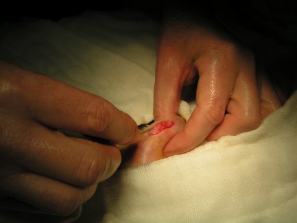
Draping maintains sterile technique at the surgical site. The scalpel is drawn along the line of the incision.
tumour_removal_08
Small tumor is being gently lifted with forceps.
tumour_removal_09
Forceps holding small tumor .
tumour_removal_10
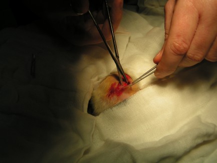
Surgeon cutting through connective tissue.
tumour_removal_11
When cutting connective tissue the surgeon must take care not to inadvertantly sever any blood vessels supplying the tumor.
tumour_removal_12
As the blood vessels are located they are clamped to prevent bleeding.
tumour_removal_13
The 2.5 cm tumor removed from the left thoracic area. Gross appearance is that of a mammary fibroma. Histopathology was declined by owner.
tumour_removal_14
Wound closure and suturing of surgical site where the tumor was removed.
tumour_removal_15
Suturing completed.
tumour_removal_16
Surgical site of large tumor prepped with an approved betadine surgical scrub, and minimal alcohol wipe.
tumour_removal_17
With the draping in place, the surgeon draws the scalpel along the line of incision.
tumour_removal_18
Blunt dissection between tissue planes.
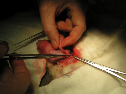
tumour_removal_19
Surgeon pulls connective tissue away from mass.
tumour_removal_20
Surgeon cutting connective tissue to resect tumor.
tumour_removal_21
Small area of cauterized blood vessel by tip of clamp.
tumour-removal-22
Clamping of blood vessel.
tumour_removal_24
Resection of tumor continues.
tumour-removal-25
Continuing to cut connective tissue.
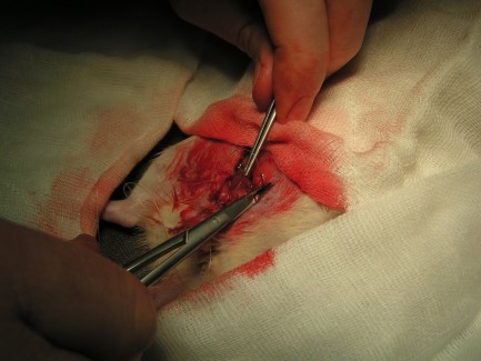
tumour_removal_26
Clamping more blood vessels. It is not unusual for large tumors to have multiple blood supplies.
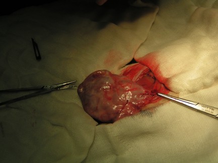
tumour_removal_27
The 5.5 cm tumor removed from the right lateral abdominal region. Gross appearance is that of a mammary adenoma. Histopathology was declined by the owner.
tumour_removal_28
Wound closure and initial suturing of site.
tumour_removal_29
Suturing of surgical site.
tumour_removal_30
Suturing completed.
tumour_removal_31
While awakening from anesthesia the rat is given oxygen.
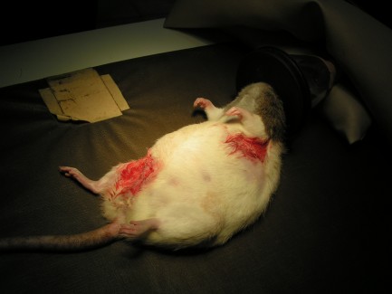
tumour_removal_32
The rat has been placed back into the induction chamber for recovery where she is provided with additional warmth and oxygen.
tumour_removal_33
She was then placed in a small hospital cage until healing was progressed enough for her to return to her usual environment.