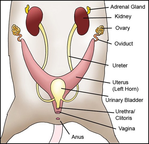
The organs of the female reproductive system are specialized to produce ova (eggs), transport the egg cells to the site of fertilization, to provide a favorable environment for developing embryos, and to move offspring outside of the body (birth) at the appropriate time.
The reproductive system also supplies nourishment for the offspring after birth and produces female sex hormones.
The main system structures of the female rat are the vagina, ovaries, uterus, and mammary glands.
Reproductive organs

Vagina
The vagina is the short muscular canal that leads from the female rat’s uterus to the outside of the body. It lies below the urethra. The vaginal walls are lined with mucous membranes which keep it protected and moist. The vagina serves as the birth canal and also as the orifice for the acceptance of sperm during mating.
When born, females have a vaginal closure membrane (vaginal plate) that typically ruptures on its own by the time the babies reach 33-42 days of age.
Ovaries
The ovaries are located at the distal end of the uterine horns near the kidneys. The oviducts connect the ovaries to each horn of the uterus. The ovaries produce the ova, also called the egg, and certain hormones.
Uterus
The uterus receives eggs and supports the growing embryos development until birth.
Rats have a uterus consisting of the right and left cornua (horns) referred to as a bicornuate uterus. This structure enables the rat to have multiple offspring.
The horns of the uterus come together to form the vagina.
Mammary Glands
Mammary glands produce milk/colostrum for the development and nourishment of young.
Mammary glands are made up of alveoli, or hollow cavities, lined with milk producing epithelial cells. The alveoli cluster in groups called lobules. The milk/colostrum drains from the lobules to the lactiferous ducts that lead to holes in the nipples.
The nipples and mammary glands can occur anywhere along two parallel lines running on the front (abdomen) side, called milk lines.
The mammary glands are then formed along these lines with six pairs of nipples or 12 nipples (though some females only have 10 nipples), three in the pectoral and three in the abdomino-inguinal regions, approximating the average number birthed in a litter.
Cells in the mammary tissue contract and push the milk from the alveoli to the nipples. Rats display complex mammary glands that consist of multiple simple single mammary glands emptying out into one nipple. (The male rat possesses rudimentary mammary glands but not nipples.)
The development of the mammary glands takes place in several phases:
At birth the epithelium is within a fat pad and has formed a ductal tree where several ducts and lateral branches steaming from each primary duct. Even at this stage the teats are still well formed.
Between birth and puberty the gland slowly grows, causing the fat pad and epithelium to extend from the nipple towards the rat’s back.
At puberty the gland begins exponential growth and rapid development of the terminal end buds (TEB). The TEB are clusters of cells several layers thick that are situated at the ends of the lateral duct branches. This rapid growth is due to cell division or differentiation of TEB cells that continues until the epithelium reaches the outer limits of the fat pad.
Following puberty the gland becomes a differentiated resting place where the TEB cells are gone and terminal ducts with small lobules or alveolar buds are common. These alveolar buds are the precursors to the large lobuloalveolar structures used during pregnancy.
Mammary development and the production of milk are affected by hormones, growth factors and environmental agents. Also, any problems during fetal development will influence further development and may affect mammary function and milk production.
Some gene mutations, such as certain hairless mutations, can affect milk production and can cause limited or no milk to be produced. Nipple structure can also affect lactation as seen with inverted nipples.
Cervix
Each uterine horn has its own cervix. The cervixes are located where the uteri connect to the vagina. Each cervix has strong thick walls.
The opening of the cervix is very small but expands to allow birth. Its purpose is to protect the uterus.
Clitoris
The clitoris is the only part of the genital structure that connects to the urethra rather than to the vagina (as seen in humans).
It is located above the urethra and is enclosed within a prepuce along with clitoral glands. The clitoris is the female homolog of the male penis. In fact, the urethra/clitoris actually visually resembles a penis making it difficult for some people to sex rats accurately.
Preputial Glands (clitorial glands)
In the female rat, preputial glands are exocrine glands that are located near the clitoris that produce pheromones.
A pheromone is any chemical produced by a living organism that transmits a message to other members of the same species. These pheromones produced by the female preputial glands are useful for attracting the male.
References
- Simmons, K. (2005, February 21). Rat Reproductive System. Retrieved December 17, 2008, from http://io.uwinnipeg.ca/~simmons/16labman05/lb8pg8.htm.
- Suckow, M., Weisbroth, S., & Franklin, C. (2005). The Laboratory Rat, Second Edition (American College of Laboratory Animal Medicine). Toronto: Academic Press.
Female Reproductive Diagram by Joanne “Bella” Hodges


