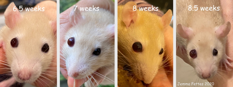
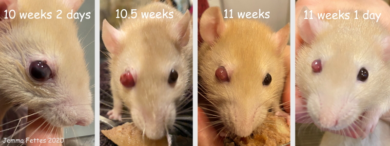
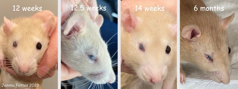
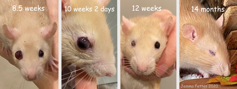
Figure 3: Buphthalmos (congenital [primary] glaucoma) in female rat (Fai).
Case history and photos
History
Fai was born a normal pup in a litter of three. She was the smallest, but not significantly so. She developed normally until 6 weeks of age.
Clinical Signs
Just after 6 weeks of age, Fai’s owner noticed that her right eye seemed to be slightly bigger than her left eye. Over the course of the next two weeks the right eye consistently grew, until it was smooth, glassy and nearly twice the diameter of the left eye. The enlarged eye was clearly irritating to Fai; she would shake her head and groom that side of her face, appearing irritated though not in pain. She also started to fall a little behind in weight gain compared to her growing siblings, despite still eating (though not as quickly as before) and being fed extras. Fai’s owner suspected that she had glaucoma.
Diagnosis
Fai was taken to see the veterinarian, who agreed that she had congenital glaucoma.
Treatment
At such a young age both the veterinarian and Fai’s owner were uncomfortable performing surgery on Fai’s affected eye. The decision was made that, as long as the eye didn’t worsen, Fai’s owner would simply observe her closely until she reached 3-4 months of age, at which point she would be better able to handle the anesthesia. In the meantime, Fai’s owner would supplement feed her and check that she was still able to blink and close the affected eye. Lubricant eye drops were prescribed in case Fai struggled with closing the eye, or the eye surface grew dry.
At 10 weeks of age the surface of the affected eye ulcerated, likely from a minor scratch. Interestingly, as a result Fai appeared to be noticeably less bothered by her affected eye. To buy time, the veterinarian prescribed the topical eye gel Isathal (containing the antibiotic fusidic acid). Initially, the injury worsened. However, after just over a week the eye seemed to be shrinking.
Over the next week the affected eye went from large and ulcerated, to a small and ultimately stable dead eye. There were now no longer any signs that Fai’s right eye bothered her at all; the pressure had clearly been relieved. Fai had been booked in for eye removal surgery once she reached 3 months of age, but after several checks the veterinarian agreed that the eye was unlikely to present any problems in the future and the surgery was cancelled a few days before the scheduled date.
Outcome
Fai is an active and affectionate girl. Her right eye has been stable and there have been no further complications. While she is clearly completely blind in her right eye, this doesn’t stop her from keeping up with her siblings. There has been no need to adapt the cage for her.
Note that, although congenital glaucoma can present bilaterally, the veterinarian does not expect Fai to develop problems with her remaining healthy left eye, as they would have been expected to show up at the same age as the affected eye.
Photos
 Row 01: These photographs show the swelling of Fai’s right eye, from the moment it was discovered by her owner at around 6.5 weeks of age, to the veterinarian’s diagnosis of congenital glaucoma at around 8.5 weeks of age. In two weeks time the affected eye had doubled in size and was clearly irritating Fai. Row 01: These photographs show the swelling of Fai’s right eye, from the moment it was discovered by her owner at around 6.5 weeks of age, to the veterinarian’s diagnosis of congenital glaucoma at around 8.5 weeks of age. In two weeks time the affected eye had doubled in size and was clearly irritating Fai. |
 Row 02: These photographs show the initial ulceration of Fai’s swollen right eye at around 10 weeks of age, and the subsequent shrinkage of the eye. During this time the topical eye gel Isathal was applied. Row 02: These photographs show the initial ulceration of Fai’s swollen right eye at around 10 weeks of age, and the subsequent shrinkage of the eye. During this time the topical eye gel Isathal was applied. |
 Row 03: Fai had been scheduled to undergo eye removal surgery at 3 months (12 weeks) of age. However, after several checks by the veterinarian the affected right eye was deemed stable and unlikely to present any problems in the future, and the surgery was cancelled. The following photographs show that there were no further changes in the eye as Fai aged. Row 03: Fai had been scheduled to undergo eye removal surgery at 3 months (12 weeks) of age. However, after several checks by the veterinarian the affected right eye was deemed stable and unlikely to present any problems in the future, and the surgery was cancelled. The following photographs show that there were no further changes in the eye as Fai aged. |
 Row 04: This row provides a quick overview of the development of Fai’s right eye. The first photograph was taken around the time that Fai was first taken in to see the veterinarian, who diagnosed her with congenital glaucoma. Due to Fai’s young age an eye removal surgery was postponed. The second photograph was taken around the time the affected eye ulcerated; the topical eye gel Isathal was prescribed. Around the time the third photograph was taken Fai’s right eye had shrunk and become stable; the veterinarian concluded it was unlikely to present any problems in the future and cancelled the eye removal surgery. The final photograph shows that there were no further changes in the affected eye. Row 04: This row provides a quick overview of the development of Fai’s right eye. The first photograph was taken around the time that Fai was first taken in to see the veterinarian, who diagnosed her with congenital glaucoma. Due to Fai’s young age an eye removal surgery was postponed. The second photograph was taken around the time the affected eye ulcerated; the topical eye gel Isathal was prescribed. Around the time the third photograph was taken Fai’s right eye had shrunk and become stable; the veterinarian concluded it was unlikely to present any problems in the future and cancelled the eye removal surgery. The final photograph shows that there were no further changes in the affected eye. |
Note to Breeders: Research shows that while Buphthalmia (congenital glaucoma) appears to be of genetic link the mode of inheritance is not fully understood. The condition appears to be both spontaneous and rare in rats. Breeders should be aware and monitor, in the event the condition appears in any line.
Reference
Young, C., Festing, M. F., & Barnett, K. C. (1974). Buphthalmos (congenital glaucoma) in the rat. Laboratory Animals, 8(1), 21–31. https://doi.org/10.1258/002367774780943797
Case history and photos courtesy of Jemma Fettes
Photo compilation by Cyzahhe
Case compilation and editing courtesy of Karen Grant RN and Cyzahhe


