Figure 6: Mammary tumor in 2-year-old female rat (Mus).
Case history and photos
History
Mus (mock mink? intact female rat) is guesstimated to have been born in January 2006.
Clinical Signs
At approximately 2 years of age a lump is discovered in Mus’s right armpit area. It feels partly attached to the overlying skin, but loose from the underlying tissue. While Mus’s keeper decides whether or not to have it removed, it continues to grow slowly.
Diagnosis
Approximately three months later, Mus is taken to a veterinarian, who diagnoses the lump to be a mammary tumor.
Treatment
An appointment is scheduled on 27 March 2008 to remove the tumor.
Gas anesthesia and a heating mat are used. The surgery takes about 40 minutes; it goes well and the tumor is removed completely (note that histopathology is not performed on the excised tumor). The incision is closed internally with sutures and externally with Nexaband tissue adhesive.
Follow-up
On the first day post-op, Mus is given a single oral dose of Metacam (NSAID prescription painkiller, dose used: 0.2mg/kg).
After the surgery Mus’s incision appears to be closed quite neatly. However, that same evening blood and blood clots are noticed at the incision site. Part of the tissue adhesive has been removed. It is unknown whether Mus did this herself, or whether one of her cage-mates did this during a brief (semi-supervised) moment together. Since only a small (outer) part of the incision has been opened up and it is no longer bleeding, it does not seem necessary to re-glue it.
Until the incision has healed completely, Mus is given Enrofloxoral Drops (antibiotic, dose used: 10mg/kg) twice a day. In addition, Dermiel (honey) wound cream is applied to the open part of the incision 2-3 times a day. After its application, Mus is distracted for 10 minutes to allow the cream time to “soak into” the wound.
Outcome
The incision heals very well, within 13 days.
Unfortunately, by July 2008 Mus has developed another mammary tumor in her right armpit area, as well as one in her left groin area. Mus’s keeper chooses not to have them removed. By September the groin area tumor has enlarged considerably, the skin has become taught, and Mus is starting to “slow down” (note that she is also becoming older). Mus is euthanized on 2 October 2008.
Photos
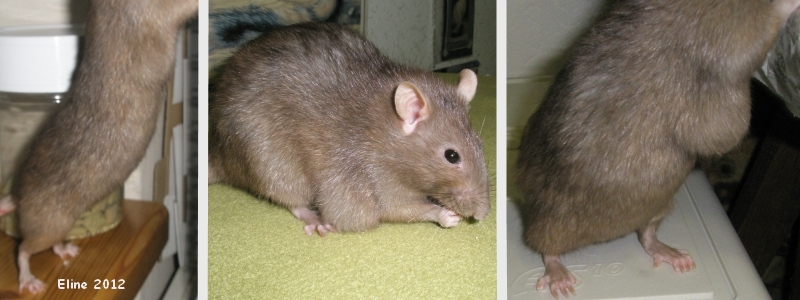 The photographs in this first row show the development of the tumor from, respectively, mid-January to early and late March 2008. |
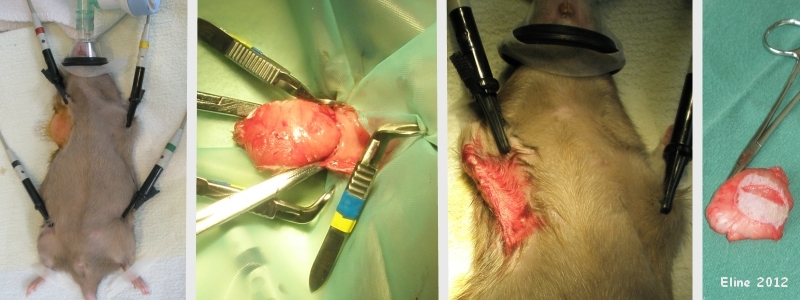 The photographs in this second row were taken during the surgery. They show the removal of the mammary tumor, with some skin attached. The shaved area of skin is yellowish due to the application of iodine (antiseptic). The black clamps monitor Mus’s heart rate. |
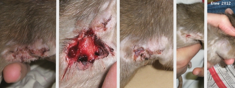 The first photograph in this row was taken shortly after the surgery; the second was taken in the evening, showing the removal of the tissue adhesive and bleeding. The third photograph was taken the following day, in the morning. The final photographs were taken, respectively, on days 7 and 13 post-op. |
Pet keeper: Eline
Case history and photos courtesy of Cyzahhe

