Figure 8: Fibrosarcoma of the leg in a 9-month-old female rat (Victoria).
Case history, photos and video
History
Victoria is a black hooded intact female rat. She and her sister were brought to a rat rescue in November 2010, at approximately 3 months of age. They went into foster care and were eventually adopted by their foster home.
The rat group is housed in a large bird cage: floor surface 60x70cm and height 150cm. In addition, for several hours a day the rats have free access to the fully enriched living room annex kitchen. Victoria is highly active, both outside their cage (e.g., climbing up into the bookshelves) and inside their cage (e.g., running in their wheel).
Clinical Signs
In May 2011, at the age of 9 months, a slightly raised reddish area develops on Victoria’s right thigh. It does not appear to bother her. Initially, the area is assumed to be an abscess and it is treated with a spray: Pyrogenium C30 + Triticum Aestivum, by Sprouting Health Ltd.
However, when the area enlarges instead of going away and another rat keeper says it looks like “skin cancer”, Victoria is taken to a veterinarian.
Diagnosis
The veterinary oncologist performs a fine needle biopsy and studies it under a microscope. Her (visual) diagnosis is sarcoma. As this type of cancer is very aggressive, immediate removal of the affected area is advised.
Treatment
Victoria undergoes surgery the following day (20 May 2011).
Afterwards, the veterinarian states that she was able to remove the affected area, but only just. This type of cancer actually requires more radical sectioning: not only of affected tissue, but also a “safe margin” consisting of healthy tissue. In this case, given the location of the affected area, this would mean amputation of the entire right leg. The veterinarian advises to have this done, preferably as soon as possible, in the hope that metastasis of the cancer has not yet occurred. However, while the veterinarian is confident that the surgery will be a technical success, she questions the effect the amputation will have on Victoria’s quality of life. She suggests to Victoria’s keeper that she contact other rat keepers and ask their opinion of how their rats managed following amputation.
Given the information, Victoria’s keeper is apprehensive regarding the invasiveness of the proposed procedure. She is also concerned over Victoria’s ability to cope with having only three legs, especially given her fondness for running and climbing about, and the fact that climbing is required to navigate their home cage and free-range area. Furthermore, it is uncertain whether or not the cancer has already metastasized. Due to this dilemma, and while waiting for the lab results to arrive (this took three weeks), the decision to amputate is postponed (note that this inevitably increases the likelihood of metastasis occurring). In the meantime, the incision heals well and Victoria’s keeper consults with other rat keepers, whose rats navigated very well after limb amputation. A decision is made: based on the fact that Victoria is a young and lively rat who is in otherwise good health, and that the amputation is not expected to have a negative impact on her quality of life, she is given the benefit of the doubt regarding whether metastasis has already taken place or not, and another surgery is scheduled.
On 28 June 2011 Victoria’s right leg is amputated, up to and including the hip joint.
Follow up
Biopsy
MACROSCOPIC
C1.1 The tissue consists of furred skin with centrally, at the level of the dermis, a moderately distinct 1.0×0.4cm mass, consisting of a network of large plump oval cells, with large oval nuclei with finely grained chromatine and a small amount of indistinct basofile cytoplasm.
The cells are surrounded by a small amount of collagen, small capillaries and foci of acute bleeding. Mitoses are 0-2PHP (MI:16).
Immunology: the tumoral cells are negative for CD13, an endothelial marker; the regional blood vessels have a distinctly positive colour (internal check).
CONCLUSION
Sarcoma, intermediate malignancy. Based on immunohistochemistry and morphology most likely fibrosarcoma. The tumor has only just (<0.1cm edge) been completely removed.
Outcome
Victoria recovers well from the invasive surgery. The incision heals within a month.
Interestingly, in order to prevent post-op stitch removal, it is regular practice at this veterinary clinic to “file down” rat incisors after surgery, to a degree that makes them considerably less sharp, but still allows the rat to eat hard foods, albeit slowly. This practice is also applied to Victoria, who does not appear to have too much difficulty eating post-op. However, she clearly prefers eating soft foods, so her keeper provides her with additional soft foods to insure that she receives necessary nutrients. Victoria grooms herself, but does not bother her stitches. Her teeth return to their normal state within 1-2 weeks.
The hospital cage is situated within the free-range area (aka living room) and is located close to their home cage. Victoria is a highly active rat, who was accustomed to prolonged free-ranging prior to her surgery. In fact, she already (successfully!) attempted to walk and climb immediately post-op! As a result, the “confinement” is increasingly stressful. Victoria’s keeper decides that it is in Victoria’s best interest to return her to their home cage. She also allows her to free-range again quite soon after the surgery.
As a result, Victoria resumes her pre-op (very high) activity level before her wound has fully healed, and without giving her body time to adjust to the missing limb. It is suspected that this lead to incorrect body movements, which possibly constricted certain nerves, as there were two episodes following the surgery during which Victoria appeared to be a considerable pain:
- A few days after the surgery, during free-range time, Victoria suddenly starts screaming. She is found lying on her side on the floor, making convulsive/spastic movements, with her head going in the direction of the wound area. Victoria’s keeper picks her up and returns her to the home cage, after which Victoria calms down.
- The second episode takes place a few weeks post-op. Early in the morning, screaming and noises are heard from the home cage. Victoria is found lying at the bottom of the cage, next to wheel. She appears to be suffering from seizures. Her sister is looking on intently. When Victoria’s keeper attempts to pick Victoria up, she is almost bitten. When Victoria’s sister approaches her, she too is almost attacked.
Eventually Victoria is placed into the hospital cage. However, she continues screaming and making convulsive/spastic movements. The veterinarian is called and a painkiller is administered. In total, the screaming lasts about 15 minutes.
Following the second episode, Victoria is taken to the veterinarian for a check-up. However, she cannot find anything wrong with Victoria and the wound has healed well. The veterinarian advises restricting Victoria’s movement for a few months. The wheel is removed from their home cage and Victoria’s free-range time is limited. Slowly, the wheel is reintroduced and the free-range time increased. No further episodes have occurred since.
All in all, Victoria required little to no adjustment to moving about with three legs. She currently runs and climbs as well as she did before the amputation. There is no decrease in her activity level.
Victoria’s only noticeable handicap is that she can no longer scratch her right side/face/ear. Interestingly, she does make the accompanying body/hip movements, so whenever her keeper notices this, she tries to scratch Victoria’s itch for her. At first Victoria would not allow this, but she has become tolerant of the scratching.
Another noticeable difference is that when Victoria eats, she does this with one hand, since the other is required to maintain her balance while sitting. However, this does not appear to be a problem for her.
Photos
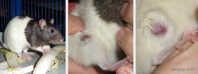 The first photograph shows a healthy Victoria in January 2011. The subsequent photographs were taken on 14 May 2011 and show the developing leg tumor. Note that the fur has been made wet to obtain a better view. |
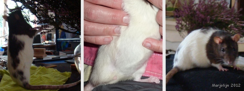 These photographs were taken in May 2012, several months after the successful leg amputation. They show the perfectly healed wound area. Note how Victoria now uses her tail for balance: it is placed in the position of the missing hind limb. |
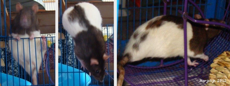 These photographs were also taken in May 2012. They are somewhat blurry, but clearly show that Victoria has suffered no loss in mobility. She still runs in her wheel and climbs about as fast as she did before the leg amputation. |
Video
Veterinarian surgeon: Ruth Fortrie
Veterinarian pathologist: Dr. Hilde De Cock
Pet keeper: Marjolijn
Case history, photos and video courtesy of Marjolijn and Cyzahhe
NL

