Figure 7: Mammary tumor removal with dehiscence in female rat (Merel).
Case history and photos
History
Merel (PEW intact female rat) adopted by present owner is guesstimated to have been born in January 2006. Familial history is unknown, although she exhibited cage territorial behavior. Merel was treated successfully for lice approximately a year prior to removal of mammary tumor. No other history of illness.
Clinical Signs
At approximately 2 years and 2 months of age a lump is discovered in Merel’s left groin area. It feels attached to the underlying tissue; the overlying skin can be moved about freely.
Diagnosis
Merel is taken to a veterinarian, who diagnoses the lump to be a mammary tumor.
Treatment
An appointment is scheduled on 27 March 2008 to remove the tumor.
Gas anesthesia and a heating mat are used. The surgery takes about 45 minutes; it goes well and the tumor is removed completely (note that histopathology is not performed on the excised tumor, but it clearly contains a lot of abscesses and pus, which – according to the vet – could be due to its fairly rapid growth rate). The incision is closed internally with stitches and externally with (colorless) Nexaband tissue adhesive.
Follow-up
Three hours post-op Merel’s incision appears to be closed very neatly. However, when the incision site is gently touched, it becomes apparent that most of the tissue adhesive has been removed, and that the skin edges are in fact no longer glued together. The incision is re-glued (using green-colored tissue adhesive). In addition, an attempt is made at bandaging Merel so that she can no longer interfere with the incision; unfortunately, this is ineffective: she wriggles out of the bandage within seconds.
Later that evening Merel removes the entire tissue adhesive, as well as all of the stitches. The result is a gaping wound into her groin area, showing the abdominal wall, muscles, and part of her leg. Two veterinarians are consulted, who offer to either re-glue the wound, or “trim” the wound edges (creating “fresh tissue”) and close it using sutures. Merel’s caretaker is concerned that re-gluing will be ineffective, and that trimming the skin will only increase the tension once sutured, causing Merel to remove the sutures as she has the glue. The decision is made to leave the wound as it is and let it heal on its own. (Note, however, that had an effective bandaging technique been available, the decision would have been to suture the incision.)
The following morning the wound appears even larger, due to the surrounding tissue having become swollen and “hard”. (This swelling remains present until post-op day 13.)
Due to the size and location of the wound, Merel is placed on her own in a one-level hospital cage. She does not appear to mind being alone; when her cage-mates are placed into the hospital cage for a brief visit they are very active, which Merel does not seem to appreciate. The hospital cage contains only fabrics and tissues, and an easy-access shoebox house with a SnuggleSafe® (heating disk) on top for warmth. The cage is kept meticulously clean.
Although Merel is slightly over-weight and there is no immediate fear of weight loss, she does seem less keen on eating than usual, so she is given porridge and baby food in addition to her regular dry mix diet to ensure that she continues eating and receives enough fluids.
For the first eight days post-op Merel is given oral Metacam (NSAID prescription painkiller, dose used: 0.2mg/kg) once a day. In addition, she is given Enrofloxoral Drops* (antibiotic, dose used: 10mg/kg) twice a day, for 14 days. Dermiel (honey) wound cream is “packed into” to the wound 2-3 times a day until it has healed completely. After its application, Merel is distracted (cuddled) for 10 minutes to allow the cream time to be absorbed into the wound. She does not object to the cream application and visibly relaxes during the cuddle sessions.
* Note: that the antibiotic was already prescribed by the veterinarian prior to the dehiscence; due to the many abscesses that were found in and on the tumor there was an increased risk of infection (e.g., pus coming in contact with healthy tissue during the surgery).
Outcome
The large gaping wound heals remarkably well, within 21 days.
Unfortunately, about six weeks after her full recovery, Merel suddenly shows signs of illness: she is weak and displays piloerection (fluffed fur). Although she is given injectable antibiotics, her condition progressively worsens over the following days and she appears to “swell up” (full-body fluid retention?). Merel is euthanized on 9 June 2008.
Photos
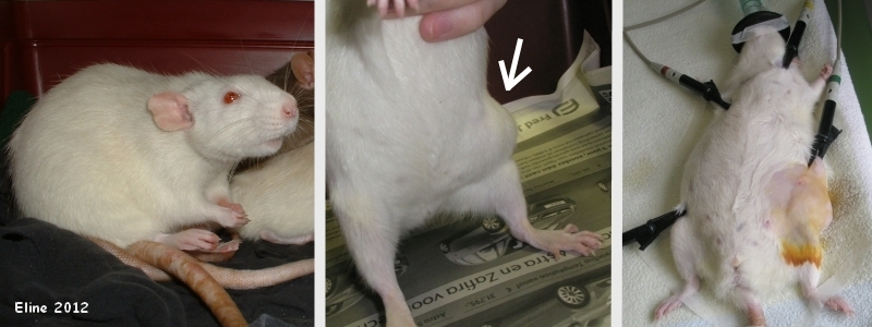 These photographs were taken before and at the start of the surgery. The shaved area of skin is yellowish due to the application of iodine (antiseptic). The black clamps monitor Merel’s heart rate. |
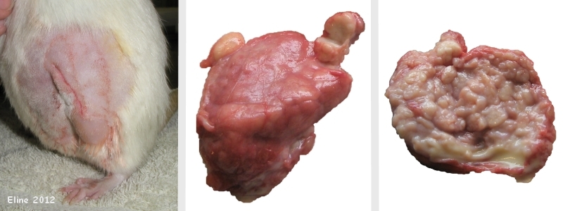 The first photograph shows the incision site three hours post-op (note: although it appears closed, the skin is in fact no longer glued together; see under Follow-up section for more information). The other photographs show the tumor: on the left the exterior of the tumor, showing an “abscess bubble” (top left) and a yellowish “tissue bubble” that has been cut transversely (top right); on the right the interior of the tumor, after it has been cut longitudinally and spread open, showing pus drainage at the bottom. |
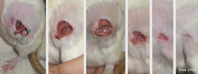 The first photograph was taken the morning after the surgery; the abdominal wall (purplish) can be seen inside the open incision, as well as a muscle (reddish) and the fascia covering the knee (greyish). The green areas on the skin are tissue adhesive. The second photograph was taken on day 5 post-op; granulation tissue has been forming inside the wound and it is slowly closing. Note that a hard swelling surrounds the wound: the skin is bulging and taut. The third photograph was taken on day 6 post-op; honey cream has just been packed into the wound and given time to melt, giving the area a shiny appearance. The following photographs show progressive healing and closing of the wound; they were taken, respectively, on days 9, 13 and 19 post-op. |
Pet keeper: Eline
Case history and photos courtesy of Cyzahhe

