Figure 3e: Obstructive Megacolon, formation of coprolith, in 30-month-old female rat (Tilty)
Case history and photos
History
Tilty: an intact, 30-month-old, fawn hooded female rat weighing 10.6 oz. (301 g). The owner procured the rat, approximated to be 2 years old, from a rescue facility. Tilty had permanent “head tilt”
Clinical Signs
One month prior to death the rat appeared to be gaining weight although the owner noted that she felt “bony” along her spine. A few days before euthanasia the owner noticed that the abdomen had become greatly distended. Within a day the rat presented with rough fur, porphyrin staining on her face, and her abdomen was rock hard.
Diagnosis
Megacolon due to unknown cause(s).
Treatment
Condition was advanced and rapidly deteriorating. Euthanasia was performed.
Follow-up
NECROPSY
The necropsy was performed immediately after euthanasia. The rat had a general odor of sepsis (foul odor from systemic bacterial infection). The rib cage was stretched out from the pressure of the mass within the abdominal cavity. A mass was noted in the colon. A lesion was noted on the colon in the same area of the mass.
The colon/cecum was removed and rinsed and a large unattached hard mass was removed. The mass was determined to be a coprolith (a hard mass of fecal matter also known as a fecaloma or a fecalith). No foreign matter was found within the fecal mass.
Lower GI tract: 5.3 oz. (149 g)
Coprolith: 1.3 oz. (38 g)
Photos
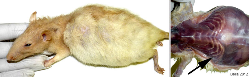 The photo on the left shows severe abdominal distention. The arrow in the second photo points to the rib cage, which had been stretched and greatly deformed by the pressure of the mass. |
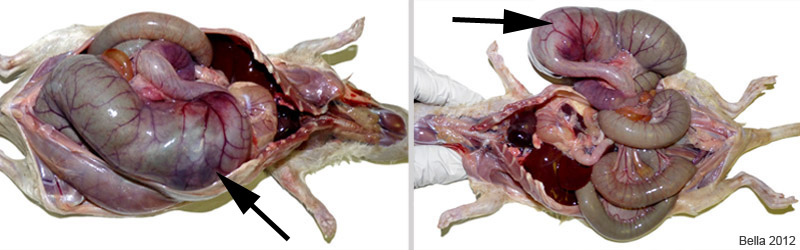 The photos above show the distended colon in situ. The arrow points to the actual location of the coprolith. The remainder of the megacolon is a result of trapped fecal matter. |
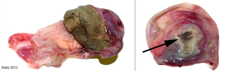 On the left you can see the coprolith. The colon/cecum has been incised and the loose fecal matter has been flushed out. After the removal of the coprolith, in the right photo, the wall of the colon is exposed. The arrow points to a severe ulceration. This lesion is most likely responsible for the systemic bacteria. |
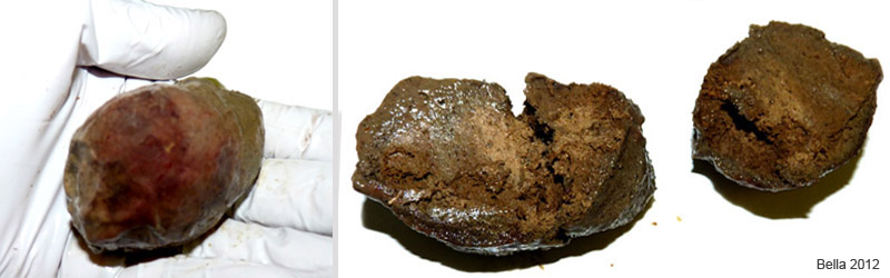 The coprolith weighed 38 grans and would have caused the rats death by systemic bacterial infection, had the rat not been humanely euthanized. |
Necropsy and photos courtesy of J. “Bella” Hodges.

