Figure 8a: Mammary tumor removal and subsequent abscess in female rat (Muys).
Case history and photos
History
Muys, a.k.a. “Muyseline” (American blue Irish dumbo intact female rat), is born on 20 July 2004
Clinical Signs
At 1 year and 3 months of age a lump is discovered in Muys’s right axilla (armpit area).
Diagnosis
Mammary tumor
Treatment
A veterinary appointment is scheduled to remove the tumor.
Gas anesthesia is used. The surgery goes well and the tumor (2.5×1.5x1cm) is removed completely and appears to be benign (note that histopathology is not performed on the excised tumor). The incision is closed internally with sutures and externally with Histoacryl tissue adhesive.
Carprofen Drops (NSAID prescription painkiller, dose used 2mg/kg) was prescribed and given to Muys as a daily single oral dose on the first, second and fourth post-op days.
Follow-up
Upon waking from surgery, Muys fairly quickly removes most of the tissue adhesive, causing the incision to bleed (note that the veterinarian was not accustomed to using tissue adhesive, but did so at the keeper’s request). Bandaging attempts are ineffective. Since the incision remains closed, the tissue adhesive is not reapplied. During the healing process Muys continues to remove remains of the glue, as well as scabs that form, thus opening up the incision site. However, it does not bleed as it did initially, and she appears to be keeping the area clean.
A fairly large portion of skin on Muys right side was shaved preoperatively. On day 12 post-op an odd circular blueish coloration appears within this area. As it does not seem to bother Muys, the veterinarian is not consulted. Over the course of the next few days it becomes clear that it is simply the first location in which the regrowth of her fur is occurring.
On day 18 post-op the nipple closest to the ventral part of the incision appears irritated: it is dark pink and slightly swollen. However, it does not seem to bother Muys, so the veterinarian is not consulted.
On day 45 post-op a soft lump is noticed in Muys’s throat area. In addition, a hard lump is noticed in her armpit area. Note that, due to its location, the latter lump may have gone unnoticed for 1-2 weeks; it is thought to be related to the nipple irritation noticed previously. Palpating the lumps seems to make Muys uncomfortable.
Although the “throat lump” shrinks noticeably in size over the course of six hours, the veterinarian is consulted the next day. The “throat lump” is diagnosed as a seroma or hematoma (probably the latter, as the area appears quite dark underneath the skin, due to blood pooling). Its cause is unclear. The “armpit lump” is diagnosed as an abscess, possibly in reaction to the dissolving sutures or glue, or a recurrence of the mammary tumor. The advice is to observe how the lumps develop.
Approximately two weeks later, the “throat lump” has dissolved completely and the “armpit lump” proved to be an abscess: it opened up and healed (Muys cleaned it without help from her keeper).
Outcome
The initial incision heals well (approximately within a month) and the subsequent abscess resolves itself (approximately within two weeks). However, it takes at least three months for Muys’s fur to regrow completely.
Unfortunately, Muys developed more mammary tumors during the course of her life (See: Mammary Tumor Figure 8b) .
Photos
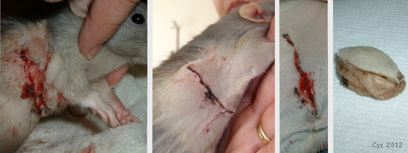 The photograph on the left was taken several hours after the mammary tumor removal surgery; the bleeding is due to Muys’s removal of the tissue adhesive. The second photograph was taken the following afternoon; note that the length and direction of the incision, combined with the way the tissue adhesive was used, has caused the skin to lose some of its suppleness. The third photograph, taken on day 6 post-op, shows continued removal by Muys of the tissue adhesive and scabs. The photograph on the right shows the excised tumor, attached to (shaved) skin. |
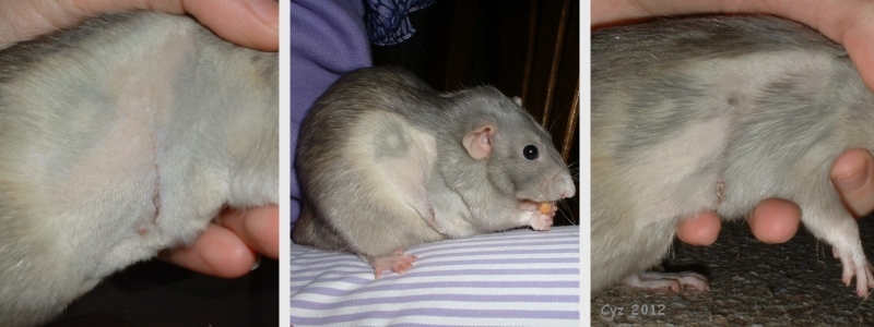 Photographs taken, respectively, on day 13, 16 and 22 post-op, showing the odd way in which Muys’s shaved fur regrew. |
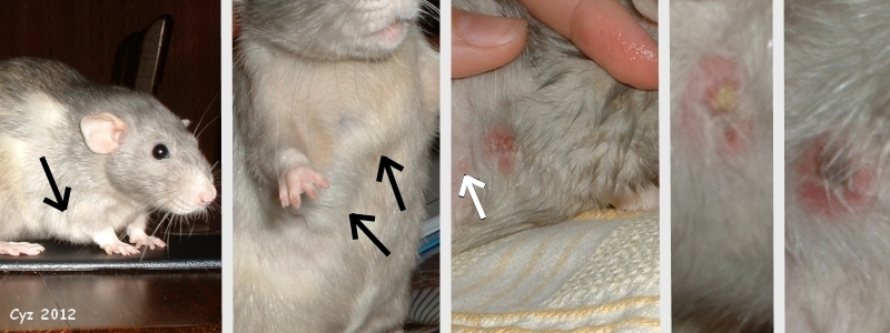 The first two photographs, taken on day 45 post-op, show the throat and armpit lumps (black arrows). The third photograph was taken on day 48 post-op (note that Muys’s fur was wetted prior to taking the photograph); it shows the abscess that is starting to scab, as well as the nipple closest to the incision (white arrow). The final photographs, taken respectively on day 50 & 51 post-op, show progressive scabbing of the abscess; it opened up on the following day. |
Case history and photos courtesy of Cyzahhe

