Figure 8b: Mammary tumor removal, postop abscess and subsequent mammary tumors in female rat (Muys).
Case history and photos
History
Muys, a.k.a.“Muyseline” (American blue Irish dumbo intact female rat), was born on 20 July 2004. At 1 year and 3 months of age a mammary tumor was surgically removed from Muys’s right armpit area (See: Mammary Tumor Figure 8a). She developed a post-op abscess and hematoma, but healed well.
Clinical Signs
At 1 year and 11 months of age Muys develops another lump, this time in her right groin area.
Diagnosis
Mammary tumor.
Treatment
A veterinary appointment is scheduled to remove the tumor.
Gas anesthesia is used. The surgery goes well and the tumor is removed (note that histopathology is not performed on the excised tumor). The incision is closed internally with sutures and despite the previous experience during which Muys removed most of it very quickly post-op the vet elected to try again closing externally with Histoacryl tissue adhesive.
Follow-up
Unfortunately, the incision site is not adhered well externally and by the following afternoon Muys has removed all of the tissue adhesive. As the internal sutures keep the incision closed, the tissue adhesive is not reapplied.
Despite the fact that the “hospital cage” is kept very clean and that it contains only “string-free” fabrics, Muys quickly develops a small (open) abscess: an off-white/yellowish discoloration can be seen inside the open incision and the incision site has a “fishy salt water” odor as described by her keeper. Muys keeps the area clean and open, allowing it to heal from the inside-out.
On day 3 post-op several “whitish flakes” are noticed near the upper perimeter of the shaved area of skin. Possibly dry skin. A few days later they are gone.
Outcome
The incision heals well within three weeks, and the abscess resolves itself within two weeks.
Unfortunately, approximately 3 months after Muys healed, the right groin area tumor returns, along with a possible recurrence of the right armpit tumor. Following an additional two months lumps also appear in Muys’s left armpit and left groin area. In addition, occasional vaginal bleeding may have indicated the presence of a uterine tumor (and/or a uterine infection).
Since there are now multiple tumors and because Muys has an accompanying respiratory problem, the decision is made not to have the tumors removed.
Especially as the right groin area tumor grows rapidly. Despite precautions* being taken against decubitus, the size of this tumor, combined with Muys being overweight and suffering from hind leg paresis (weakening), causes a pressure sore on her left knee, for which the veterinarian prescribes Metacam (painkiller) and Clindobion cream (clindamycine 10mg/kg) 2-3x/day.
-
* Precautions that should be taken against decubitus:
- provide good nutrition
- provide a thick padded layer of soft and wrinkle-free bedding/nesting material
- rotate the patient’s sleeping/sitting position every few hours
Despite all of her inconveniences (or at least until the pressure sore developed), Muys remained in fairly good spirits. However, as her respiratory infection stops responding to medication and becomes increasingly severe, with more frequent “gasping attacks”, both Muys and her keeper become tired of fighting the slow decline of Muys’s body. Muys is euthanized on 27 January 2007, at the age of 2 years and 6 months old.
In retrospect, due to the frequent “gasping attacks” and the size of the groin tumor, Muys’s keeper feels it would have been in Muys’s best interest to have her euthanized sooner, possibly two months earlier.
Photos
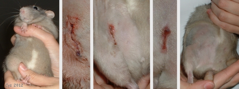 The photograph on the left, taken prior to the mammary tumor removal surgery, shows the lump in Muys’s right groin area. The second photograph shows the condition of the incision a short while after the surgery. The subsequent photographs, taken respectively on day 4, 10 and 22 post-op, show the healing process. On day 4 the slight infection is visible, which Muys kept clean; the whitish flakes can also be seen. By day 22 the incision has healed completely. |
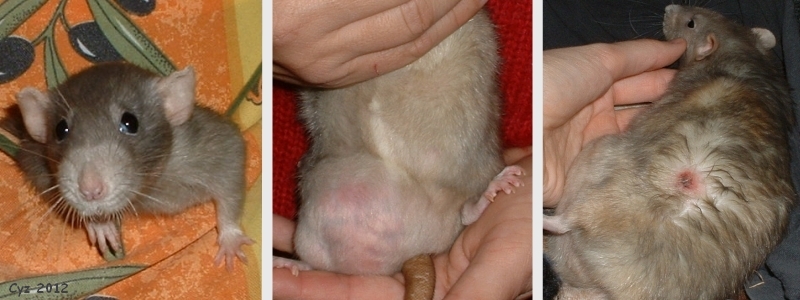 These photographs show Muys a few weeks prior to being euthanized. In the photograph on the left she is being carried around the house in the pocket of an apron. The middle photo shows Muys’s enormous groin tumor. The photo to the right shows the pressure sore it caused. |
Case history and photos courtesy of Cyzahhe

