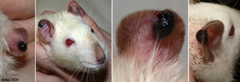
Figure 1: Melanoma in a 1-year-old male rat (Apollo).
Case history and photos
History
Apollo, a one-year-old neutered male rat.
Clinical Signs
Apollo had a small dark bulging spot on his right ear. Over a 5-month period the spot darkened, became nodular, enlarged, and bulged from both sides of the ear.
Diagnosis
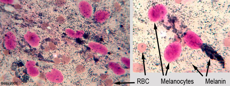
It was initially diagnosed as an injury related hematoma and the prognosis was that it would resolve itself. After 5 months the affected area had enlarged significantly. A fine needle punch biopsy was performed and the cells were examined microscopically resulting in a confirmation of melanoma..
Treatment
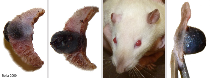
The melanoma was excised using a laser. The ear was re-modeled to be aesthetically pleasing.
Outcome
The rat recovered rapidly after the surgery. The owner was very pleased with the new small ear. She will be watching him closely for any other spots or nodules.
Photos
 The above photos show Apollo’s melanoma before it was removed. |
 These photos show the affected portion of the ear that was removed as well as Apollo’s “new” smaller ear. |
 The above photomicrographs, from the needle biopsy of the nodule, show the melanocytes (cells that produce the pigment melanin) and an abundance of melanin. |
Diagnostics: Vanessa Pisano, DVM
Surgeon: Richard McKinnis, DVM
Microphotography: J. “Bella” Hodges


